Category: Biosecience

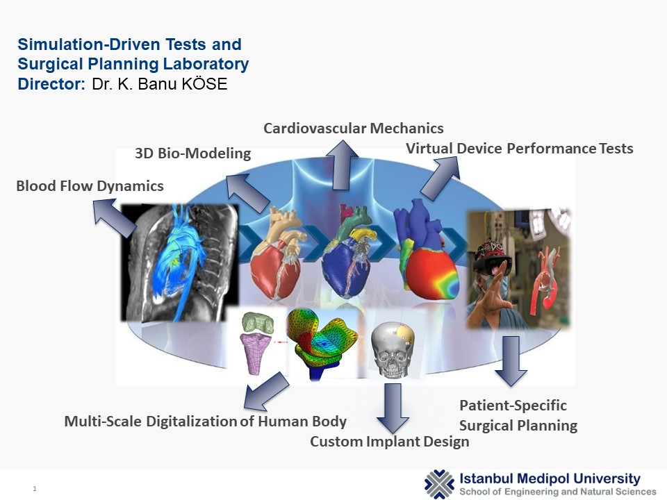
Surgical Planning Laboratory

MEDIPOL UNIVERSITY

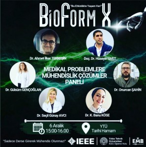
Engineering Solutions to Medical Problems

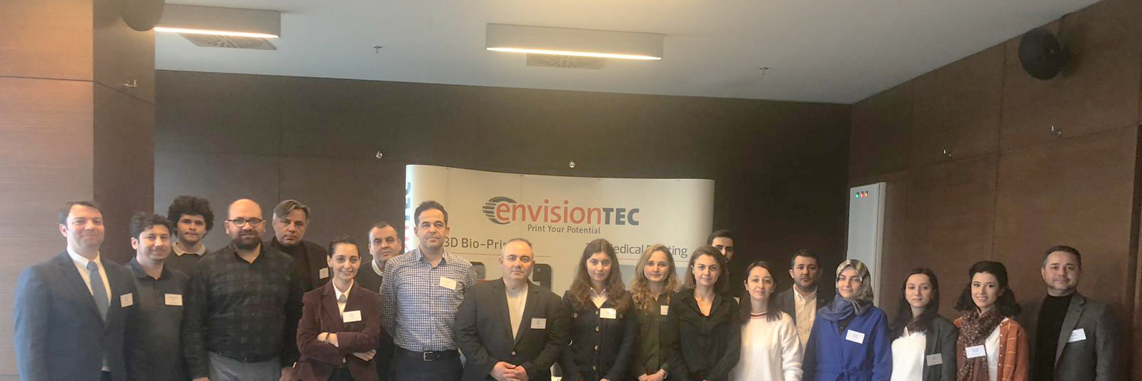
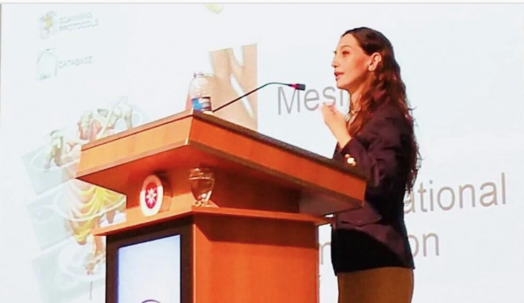
We met at the ‘Engineering Solutions to Medical Problems’ panel at Yıldız Technical University. Many thanks to ~Yıldız Technical University IEEE Engineering in Medicine and Biology Society~ for this well-planned organization.
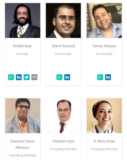
EVBio

A team I am honored to be involved in EVBio!
EVBio is a digital think tank formed by scientists from all disciplines related to vascular medicine, from molecular biology to scientific computing. Our mission is to imagine the future through disruptive basic and translational research. Our team works tirelessly to formulate one universal coherent theory for vascular disease and support all global efforts towards its conception and validation.

Our membership will increase soon!

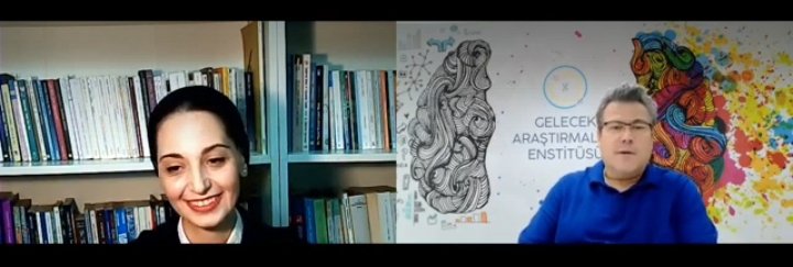
Cardiovascular Engineering in Istanbul

We talked about cardiovascular engineering and its future.   I was honored to be a  guest in Future Research Institute. Many thnaks to Coskun Dolanbay.

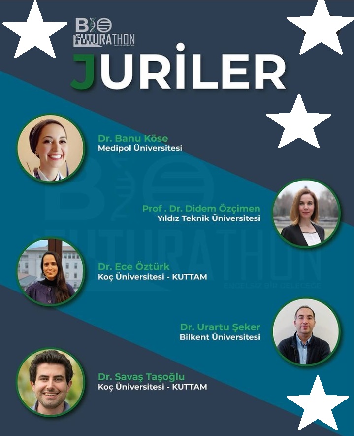
Bio Futurathon

We discussed how we can reach a ‘life without barriers’ in the future and evaluated the projects that the contestant groups developed during the event  day within the context of hackathon. Congratulations to the winning teams.

For more details : BioFuturathon and Future Research Institute

Source

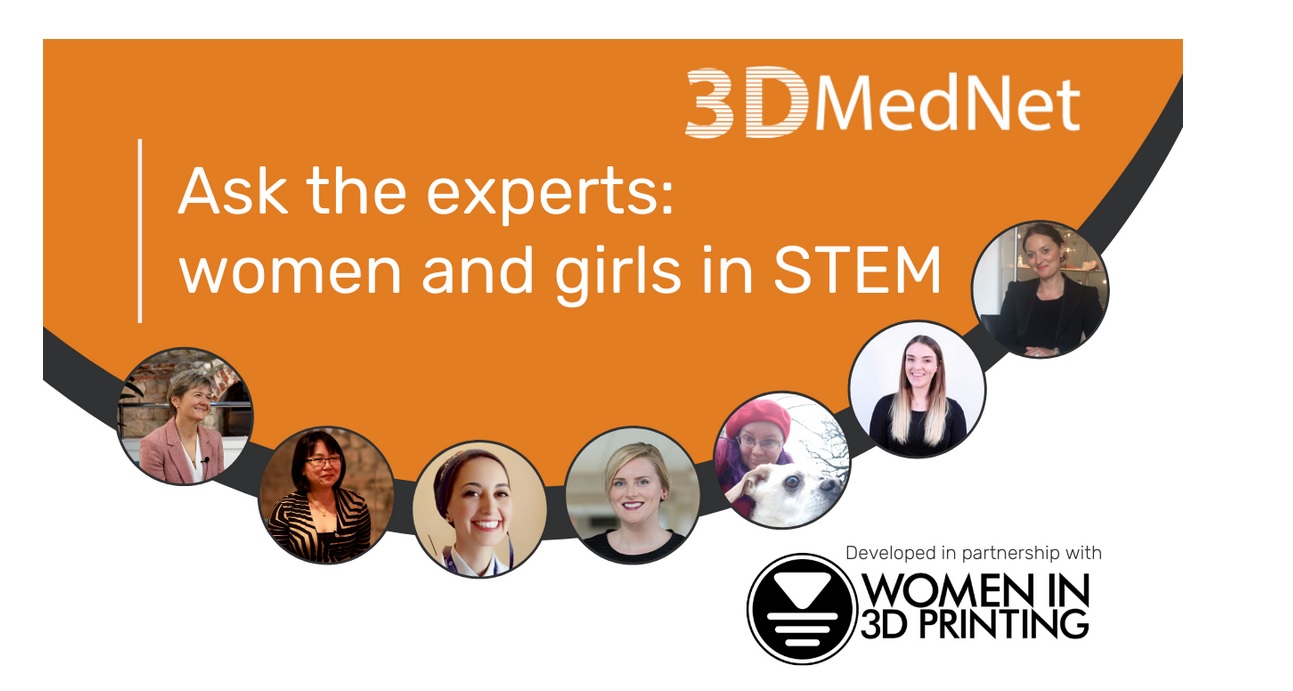
Women in STEM

Kevser Banu

To commemorate International Women’s Day (8 March 2020), 3DMedNet has put together our first ‘Ask the experts’ feature in partnership with the global organization, Women in 3D Printing.  Thanks to Georgi for inviting me to the conversation. Check the link for the full interview.

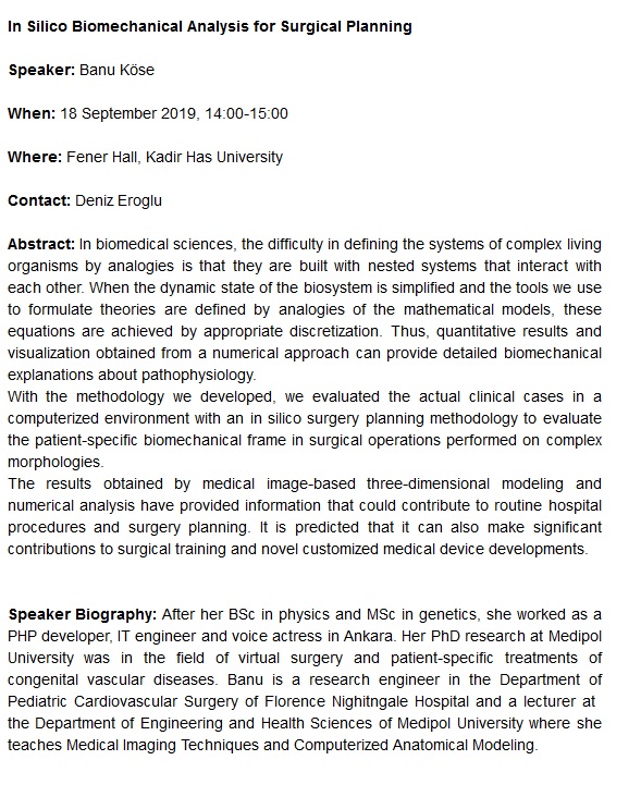
In Silico Biomechanical Analysis for Surgical Planning

Soruce: http://complexsystems.khas.edu.tr/ , https://nodds.khas.edu.tr/node/115

Women in 3D Printing

I was the guest of Women in 3D Printing this week.

The full-page is on this page: https://womenin3dprinting.com/banu-kose/

Thanks to Nora Toure for all the great work she has done and for bringing us together.

Read more »

Institute of Physics / Challenges in Cardiocascular Flow

A joint meeting showcasing current work addressing the complex challenges in cardiac flow modeling, particularly focusing on the work of early career researchers (source).

Physics

Liquid State Physics in Turkey

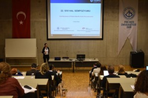
22. Liquid State Symposium (22. Sıvı Hal Senpozyumu) took place on 7th December 2018 in Piri Reis University.
It was very proud to be together with the physicist academics I knew and admired since my undergraduate years.
I find myself lucky to see the Prof. Zehra Akdeniz that I have always admired and exemplified. I could finally meet Prof. Nihat Berker who is not only a famous physicist but also an intellectual on comparative literature readings.

Thanks to Dr. Ozan Sarıyer and Dr. Gulsen Evingur for organizing this meeting.

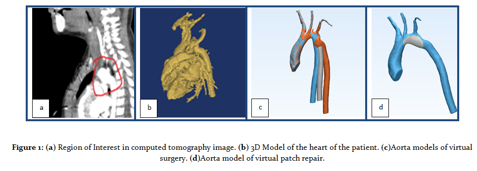
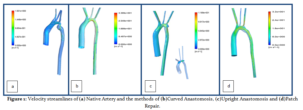
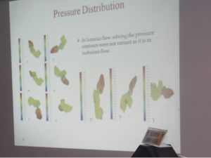
Prof Pekkan presented biological flow researches of his lab, and I presented a sample case of a pediatric aortic blood flow comparison study which is done with the great help of Dr. Ece Salihoglu.

DONE

Optimizing your workflow in the Mimics Innovation Suite

The Mimics Innovation Suite (MIS) allows you to automate your workflows, potentially saving a lot of time, achieving more consistency, and reducing repetitive work and human error. That is an easy thing to say, but if you do not have much experience with scripting, we all know that it can be tough to get started. If you want to speed up your learning curve and get a head start, then this could be an interesting training for you.

Topics will include:

Basics of Optimizing your Workflow in Mimics 21 and 3-matic 13
How to write your first scripts
Introduction to Python
Hands-on training exercises for creating planning workflows (e.g. loading datasets, performing basic segmentation steps, landmarking, creating anatomical coordinate systems, designing custom implants)

Simscale Certification

Thanks to Anna Flessner and Milad Mafi for the certificate, excellent training documents and lectures. #Simscale

The workshop was including the simulations of

1- Hip Joint Prosthesis and comparison of different materials with displacements

2- Stent Design and Comparison of different materials and different balloon pressures

3- Comparison of different stenosis persentages in carotid artery samples by CFD

Utrecht Medical Center / TGA Symposium

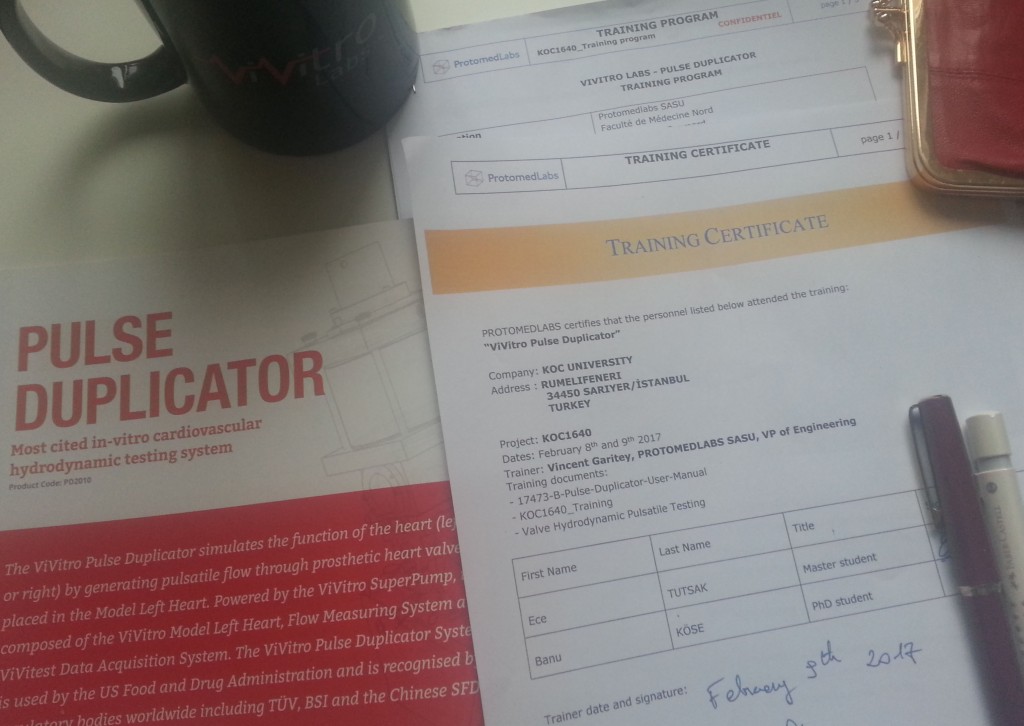
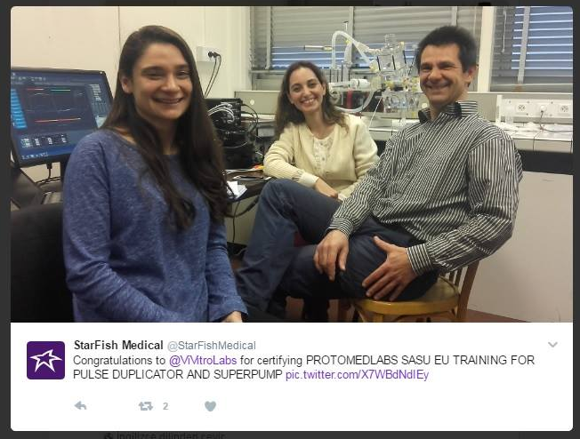
Vivitro Pulse Duplicator Training in Protomed Labs

http://www.protomedlabs.com

It was really a great experience at  Protomed Labs in Aix-Marseille University. I really enjoyed learning about hydrodynamic testing requirements, Vivitro Pulse Duplicator, its calibration, flow testing, heart valve testing, and at the same time  practicing.

Thanks to Prof. Kerem Pekkan for suggesting this training for Ece Tutsak and me.

I would like to express my sincere thanks and gratitude to  Karim Mouneimne and Vincent Garitey for all the kind care they took, regarding the training, sharing their expertise to us, the  detail notes, all the answers whenever required etc. in Protomed Labs.

I hopefully will be able to implement it further into my field.  This got me inspired and ready to go!

For More

20. National Liquid State Physics Symposium 16 December 2016

20. National Symposium on  Liquid State Physics was held in Piri Reis University.

The symposium was obtaining various studies about liquids as water and climate change, simulating strait systems, oceans, spin glass phases, liquid crystals, serum transferring,  swollen gells, GO composites, metals with  glass-like structure, super hidrophobic polistren and, biofluids {yes, this was mine ;) }.

It was an incontrovertible experience for me that i could meet new studies in the field and spend nameable times with  physics authors.

Many thanks to organizing comitee (especially to Gülşen Evingür) and Sevtap Yıldız Özbek.

The website of the symposium is here.

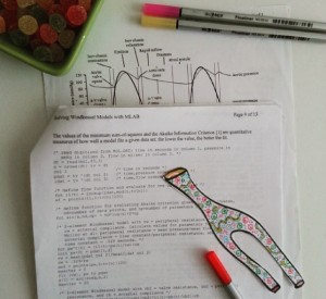
Windkessel plus colours ;)

I know why I’d rather CFD. Because it has colours. Windkessel analogy has not.

Link

Simulate the Physiology & Understand the Pathology

.

Computational Life is a young company which has the specialty on computational flow simulations and mathematical models for the medical technology field.

The validated software Digital Avatar Platform (DAP) of Computational Life is modeling human and animal body mathematically. It is testing physiological scenarios for drugs, medical devices and treatment methods.

Circulation system, cerebrospinal fluids, transport of pharmaceutical products throughout the body can be simulated for the human and animal body with DAP. It can also be modified due to the experiment.

They replied to me with a very warm and energetic mood when I wrote them. It is great that there are enthusiastic people in the medical technology field. I am sure that I will hear more about the news of Computational Life in the next days.

Thanks to Christian Contarino, Davide Chieco and Carlo Rivis for their innovative platform which brings a great help for clinicians, researchers, and engineers.

Please check their website for more information.

.

ICPT – GEFIK 2016

I had chance to present my works to authors and answer the questions of young curious physicisits at GEFIK2016 in Ege University. Discussing about medical physics and classical mechanics with physicists was a peerless experience.

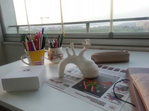
3D Printed Aorta

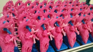
A pediatric aorta model reconstructed from the 3D CT images.

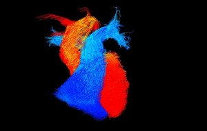
‘Go with the flow’ by Victoria Stoll

The British Heart Foundation (BHF) announced the winners of its annual ?Reflections of Research? image competition, reflecting the charity?s research into heart and circulatory disease.
The winning image ? titled ?Go with the flow,? by Victoria Stoll, a BHF-funded researcher at the University of Oxford ? captures the blood flowing within an adult heart frozen in time. Blood flows within the main pumping chambers (ventricles) of the heart and the vessels leaving the heart. The blue flow is blood that lacks oxygen and is travelling to the lungs. The red flow is blood that has been through the lungs and received oxygen and is now ready to be pumped around the body.
Stoll is using this type of imaging, four-dimensional cardiac magnetic resonance imaging (MRI), to look at the blood flow in four dimensions within the hearts of people with heart failure, whose hearts are not pumping effectively. She has already found that in people with severe heart failure the blood flows around the heart in a more disordered and disrupted pattern.

More

ISCOMS – Faculty of Medical Sciences- Groningen University

Interactive Surgical Operation

ISCOMS 2016

Workshop – Anatomy of Thef Heart with Michiel E. Erasmus MD PhD – Faculty of Medical Sciences- Groningen University

6ISCOMS 2017 at University of Groningen

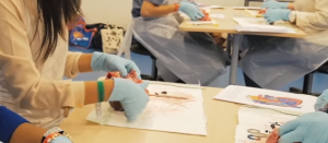
Many thanks to University Medical Center Groningen for the oral sessions and workshops of 3D Lab, LVAD treatment, Dissection of Brain, CABG treatment, IV Injections and Nuclear Medicine.

Mimics Innovation Course 2017 – Belgium

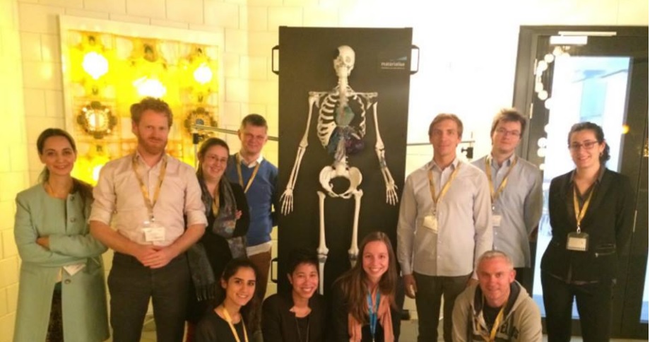
Materialise provided a Mimics Innovation Course on Soft Tissue.

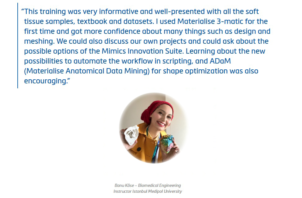
This training was very informative and well-presented with all soft tissue samples, text book and datasets.

I used 3-Matic for the first time, and got confidence about many things about design and meshing. We could also discuss our own projects and could ask possible options of Mimics Innovation Suite.

Learning about the news about scripting possibilities to automate the workflow, and ADam (Materialise Anatomical Data Mining) for shape optimisation was encouraging.

Thank you for sharing your knowledge with us, Karen de Leener and Inés da Silva.

Thank you for the great help of Job der Kinderen.

Anatomical Modeling & 3D Printing Meeting with 4C Medikal

PRINT THYSELF

This sort of procedure is becoming more and more common among doctors and medical researchers. Almost every day, I receive an e-mail from my hospital?s press office describing how yet another colleague is using a 3-D printer to create an intricately realistic surgical model?of a particular patient?s mitral valve, or finger, or optic nerve?to practice on before the actual operation. Surgeons are implanting 3-D-printed stents, prosthetics, and replacement segments of human skull. The exponents of 3-D printing contend that the technology is making manufacturing more democratic; the things we are choosing to print are becoming ever more personal and intimate. This appears to be even more true in medicine: increasingly, what we are printing is ourselves.

Source: Newyorker

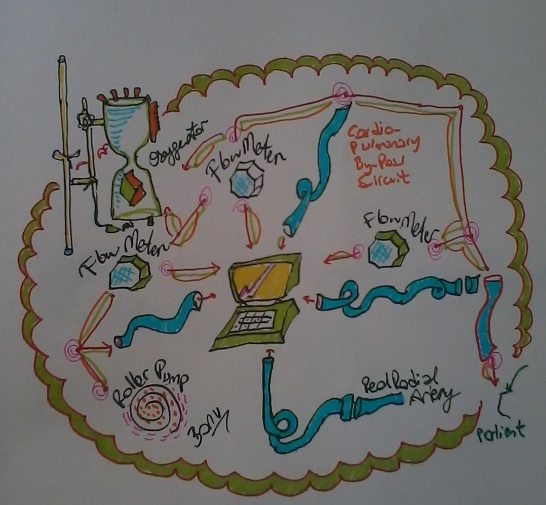
Cardio-Pulmonary By-Pass Circuit

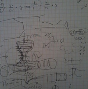
I simplify by drawing.

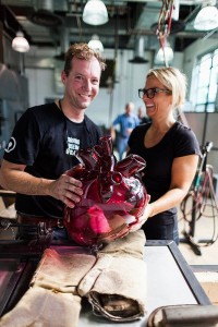
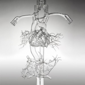
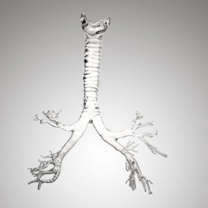
Gigantic Human Organs Made of Glass


Modeling human anatomy. Isn’t it fabulous?

Read more.

Many thanks Sıla Yavuz for this pleasant link.

Trando Med

Trando Med will attend MEDICA 2017 in the Dusseldorf Germany from 13-16 November 2017. The booth is Hall 13 Booth F 9-05

The Impacts of Cardiac MRI on Congenital Surgical Results

On October 19 Koc University presented “The Impacts of Cardiac MRI on Congenital Heart Surgical Results,” a public symposium at Koc University Hospital Artlab Conference Hall that explored the benefits of cardiac MRI scans for depicting the congenital heart anatomy.

The symposium began with a presentation by Prof. Afksendiyos Kalangos. He was followed by renowned pediatric cardiovascular surgeons and cardiologists Prof. Atıf Akçevin, Prof. Alpay Çeliker, Prof. Aphrodite Tzifa, A. Professor Tijen Alkan- Bozkaya and the radiologists Dr. Serhat Aygün, A. Professor Özdil Başkan who presented their own experiences, demonstrating a vast range of approaches to defining the context.

Prof. Kerem Pekkan presented his studies with MRI imaging and cardiovascular mechanics in the cases of blood flow dynamics, biomaterial tests, patient-specific vascular materials and surgical planning for congenital heart diseases. His signified projects were one of the fascinating parts of the symposium for participants.

Hemodyn

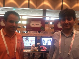
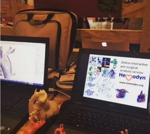
Hemodyn, the first cardiovascular mechanics and surgical planning company of Turkey is taking place in StartUp Istanbul 2015.

Hemodyn is assisting the surgeons in the diagnosis and surgery planning of the congenital heart diseases in The Incubation Office of Koc University.

Hemodyn Team has always been an invariable place for my research vision. I want to thank to Kerem Pekkan, Şenol Pişkin and Volkan Tuncay by means of this event.

Link

IFG26 – Statistical Physics Days

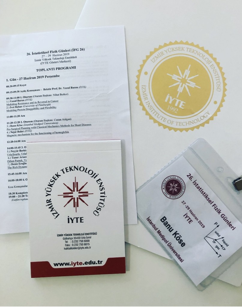
26th Statistical Physics Days were held in İzmir Institute of Technology.

During the program organization, Prof. Nejat Bulut’s dedication and careful attention to every detail was so amazing that it will be a very nice experience in my mind.

It was an honor to be among the successful physicist academicians and to listen to their work. It was also my chance that I had the opportunity to talk about my own practice and find the opportunity to discuss it with very precious professors.

K. Banu Kose

3D Printing for Pediatric Cardiothoracic Surgeons

‘Having worked in product development for the past few years, Dr. Enrique Garcia had seen what 3D printers were capable of and began investigating the possibilities for creating models for pediatric cardiologists to use before an operation. She began by asking surgeons from around the country what they thought of the idea. To say that their response was overwhelmingly positive is an understatement. The value of this idea was immediately apparent.’

‘Pediatric heart surgery is the hardest thing that I can imagine a person doing. A surgeon doesn’t know what he’s going to see until he opens a child?s chest. Every heart is different and every cardiopathy is different,? said Garcia. ?A baby?s heart is the size of a walnut, and surgeons need to go in and move around structures that are as small and thin as a human hair, and they’re doing it with their own two hands. And all of this is occurring against a ticking clock.’

‘Having something in your hands, and being able to turn it any way you want, and to be able to cut and open it up and see the inside; and to be able to physically hold it, to feel it, is something that can?t be replicated on a computer.’

Read More in the source.

cSound

Researchers have created software that can model internal organs in ‘extreme 4D’.
The system, dubbed cSound, is currently being used by cardiologist Bijoy Khandheria, who has been fixing broken hearts for more than three decades.

Dr Khandheria describes the images as ‘exquisite’, and says it’s like opening up someone’s chest and watching their heart beat.’

‘Traditionally, ultrasound has allowed us to see the heart but not in as much detail as we might like,’ he said.
‘We used the signal to image the heart layer by layer, almost like a butcher using a knife, and then mentally splice the layers together to see the whole picture’.
Dr Khandheria and his colleagues at Aurora St Luke’s Medical Center in Milwaukee, have recently started ‘extreme 4D’ software.
The images are so clear that it allows doctors to see how blood swirls around clots in arteries.
This can then be used to measure the severity of blood leakage around the valves and assess the damages.
‘It’s almost as if I took out the valve and started turning it with my hands,’ said Dr Khandheria.

Read more

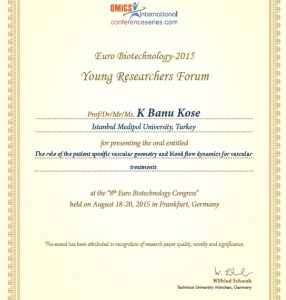
Omics Conference

3D-Printed Artificial Heart Test / #ETH Zurich

Voksel 3D Surgical Planning with Simpleware -İstanbul

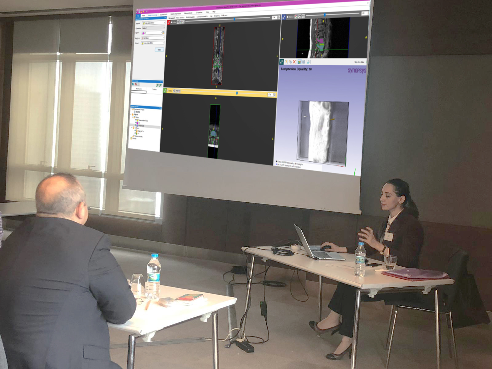
We depicted a live- surgical planning scenario with Prof. Erbil Oğuz and Kerem Girgin in Voksel 3D event. We used Simpleware for image processing, segmentation and designing.

Joint Interventional Meeting / Rome

Joint Interventional Meeting will be held on 12th February in Rome with the partipiciant TCT(Transcatheter Cardiovascular Therapeutics).

The meeting will be about the very latest Innovation on Interventional Cardiology and to approach New Techniques through educational Live Cases.

Congress directors are Antonio COLOMBO, Eberhard GRUBE, Martin B. LEON, Carlo DI MARIO, Jeffrey W. MOSES, Gregg W. STONE

Click here for the congress program.

The Office

8th Istanbul Symposium: Pediatric Cardiac Surgery in Turkey, Developing CPB & ECLS Systems and Suggestions for Decreasing Complications

The 8th Istanbul Symposium will be held at Medipol University Mega Hospital.

10 January 2015, Saturday, 9:00-18:00

Symposium Comittee:

Prof. Dr. Atıf Akçevin, Yrd. Doç. Dr. Tijen Alkan-Bozkaya, Prof. Dr. Halil Türkoğlu, Prof. Dr. Akif Ündar

Click here for attending and the booklet.

Many thanks to Mehmet Hikmet Üçışık for his kind apprising.

ECVS Conference

It was  sensible showing 3D printed models when i was presenting my study.

Carol Malnati

“- I wanted to be someone that encouraged young women to get involved in math, science, and engineering.”

Today, she’s doing just that.

As a product development engineer in the Medtronic cardiovascular division, Carol has been doing what she loves for more than 25 years. She provided critical technical expertise for the company’s first implantable cardioverter defibrillator and continues to collaborate with engineering teams and physicians to find new ways of doing things.

But on top of her day job, she has taken on another commitment – overseeing the Women in Science and Engineering (WISE) Initiative at the company.

Beginning in the spring of 2017, Medtronic introduced another opportunity that taps into an often overlooked talent pool.  Careers 2.0 is a “returnship” program designed to provide paid internships for female engineers looking to get back into STEM-related careers. Research suggests close to 25 percent of women in engineering careers leave the industry by age 30, citing work culture or family commitments.

“This is a way to bring these talented women back into our technical and managerial ranks,” says Carol. “We are very excited about providing this amazing pool of talent an opportunity at Medtronic.”

“Overall, I want to inspire women,” says Carol. “Whatever your passion is; clean air, fighting hunger, or improving healthcare. Behind the biggest challenges of humanity, there’s an engineer working to find a solution.”

Source

The special session for the women in the field of cardiovascular surgery – The 64th Istanbul ESCVS

International Congress of the European Society for Cardiovascular and Endovascular Surgery (ESCVS) will be held on March 26th – 29th, 2015 in İstanbul in collaboration with International Congress of Update Cardiology and Cardiovascular Surgery.

The congress scientific program includes a session for women in cardiovascular surgery which will be held on March 28th.

Abstract Submission Deadline
December 22, 2014
…………………………………………..
Notification of Abstract Acceptance
January 2, 2015
…………………………………………..
Early Registration
until November 7, 2014

ESCVS 2015 Web Site

Starfish Medical – VivitroLabs – ProtomedLabs – Marseille – France


Ece Tutsak (Left) – Banu Köse(Middle) – Vincent Garitey(Right)

Heart Flow

Using data from a standard CT scan, the non-invasive HeartFlow Analysis creates a personalized 3D model of the coronary arteries and analyzes the impact that blockages have on blood flow. See the website: http://www.heartflow.com/

The Horizon for Mechanical Circulatory Support

Filmed at the 2014 STS Annual Meeting in Orlando, Florida, this roundtable discussion focuses on mechanical circulatory support. John Kern moderates the discussion with Pavan Atluri and Francis Pagani. The panelists discuss mechanical circulatory support, LVAD therapy, and heart transplantation. The discussion concludes with thoughts on the future of mechanical circulatory support.

Source:  CTS

2014 AATS Cardiovascular Valve Symposium in İSTANBUL

The inaugural 2014 AATS Cardiovascular Valve Symposium will bring international leaders in adult, congenital, and adult-congenital heart valve disease as well as diseases of the ascending aorta together for three days to discuss the latest information regarding management guidelines, imaging, pathology, minimally invasive approaches, percutaneous approaches, surgical techniques, devices, and long term results. Faculty presentations of the latest available data, techniques, and state of the art reviews will be supplemented by comprehensive surgical video sessions. In addition, the program will include abstract presentations selected by the program committee from submitted original work on a wide range of topics. This innovative program will allow attendees at all levels to advance their knowledge in aortic and ascending aorta, mitral, pulmonary, and tricuspid valve disease across all age spectrums during this AATS Symposium in Istanbul.

Click here for details.

PROGRAM DIRECTORS

David H. Adams
Mount Sinai School of Medicine
New York, New York, USA

Sertac Cicek
Anadolu Medical Center
Istanbul, Turkey

Joseph S. Coselli
Baylor College of Medicine
Houston, Texas, USA

Pedro J. del Nido
Children’s Hospital
Boston, Massachusetts, USA

Clinical Engineering Lecture in Beykent University

Clinical Engineering

Lecture

ESCVS Congress 2014

VOKSEL 3D Event in Istanbul

Voksel’s Anatomical Modeling, Surgical Planning, 3D Printing with Engineer – Surgeon Collaboration Training‘ was held on 23rd February in Istanbul.

I had the chance to share my experiences in image processing and modeling with the participants. I would like to thank Kerem Girgin, Erbil Oğuz, Samet Serbest and Cansu Çeltik from Voksel. It was great to be a part of Voksel team, and meeting with the participants who were aware of the benefits of interdisciplinary collaborations and patient-specific planning very well.

Surgical Planning and 3D Printing Meeting

36th Annual Recent Advances in Clinical Nuclear Cardiology and Cardiac CT: State of the Art Updates and 101 Evidence-Based Case Reviews

The 36th Annual Recent Advances in Clinical Nuclear Cardiology and Cardiac CT: State of the Art Updates and 101 Evidence-Based Case Reviews course is filled with this information and more to meet your needs! Cardiac imaging and non-imaging cardiovascular professionals will benefit from this stimulating and interactive two-and-a-half day multidisciplinary course that explores the latest advances in nuclear cardiology, cardiac CT and other imaging modalities across the spectrum of patients routinely seen in a typical practice.

This course addresses the call for increasing quality in performing, interpreting, reporting and applying nuclear cardiology and cardiac CT studies while emphasizing physician competency and optimal laboratory performance of imaging studies in coronary artery disease and non-coronary cardiovascular disease.

If your interest is in multimodality cardiac imaging and understanding the complexities of image acquisition, interpretation and cardiac imaging while, at the same time, dealing with the scrutiny of escalated use and increased costs facing cardiac imaging, this course is designed for you.

Click for registering

3D Bio-Printing Project of Sabancı University

For the first time in the world, tissue structures were created by using self-supported live cells in a 3D bio-printer from medical images in the 3D Tissue and Organ Printing Project.

Sabancı University Faculty of Engineering and Natural Sciences? Manufacturing Systems Program professor Bahattin Koç and his stedents; Can Küçükgül, Saime Burçe Özler, Forough Hafezi printed artificial tissue construct at the Nanotechnology Research and Application Center (SUNUM) using self-supported live cells in a  3D bio-printing system.

The 3D Tissue and Organ Printing Project team used live human dermal fibroblast cells as bio-ink to print a part of aortic tissue.  Human blood vessel tissue consists of mainly three types of cells: fibroblast, endothelial  and smooth muscle.  Fibroblast cells are the main cells of connective tissues.  They synthesize the extracellular matrix and collagen protein needed for tissues.  Endothelium is the thin inner layer of cells of blood vessels.  Smooth muscle cells are found in inner organs such as blood vessels, esophagus and intestines.  The scientists continue their efforts to maturate the blood vessel tissue created by fibroblasts as well as endothelial and smooth muscle cells in a bioreactor.

Read More

3D Modeling to Display Complex Congenital Heart Diseases

Thanks to Emre ^.^

My Lab Drawings – II

A pulmonary artery. Actually this was the first step of my master thesis.

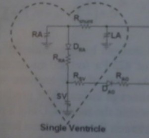
Single Ventricle Analogy

Link

17th U.S. National Congress on Theoretical & Applied Mechanics

Every four years since 1950 the leading mechanics researchers  have convened the U.S. National Congress on Theoretical and Applied Mechanics. All mechanics researchers and students are invited for the 17th  Congress on the beautiful Big Ten campus at Michigan State University. The sessions will be held at the Kellogg Hotel and Conference Center on Michigan State University’s campus.

Many thanks to Seungik Baek for his kind invite us Cardiovascuar Mechanics Minisymposia:

The goal of this minisymposium is to provide the state of the art in theoretical and computational methods applied to the cardiovascular mechanics including computational and constitutive modeling, theoretical vascular mechanics analysis, and cardiovascular design technologies. Topics may include, but are not limited to cardiovascular fluid and/or solid mechanics, cardiovascular diseases and treatment, optimization techniques. We believe that you would be an excellent contributor to this session based on your many exceptional works in the fields of theoretical and computational mechanics to cardiovascular problems.

The 14th Annual International Symposium on Congenital Heart Disease

The 14th Annual International Symposium on Congenital Heart Disease will feature a world-class faculty of domestic and international experts in Cardiology, Cardiac Critical Care, Cardiac Surgery, Nursing, Hospital Administration, and Ethics. This year the conference will focus on Diseases of the Cardiac Valves from the Fetus to the Adult. The program will include didactic, case-based, and interactive presentations as well as pathologic heart specimens and practical workshops. Special tracks dedicated to cardiovascular nursing and hospital administration will be included making this a truly team-based symposium.

Click for the Symposium Web Page.

Click for Symposium Document.

The World Congress of Biomechanics (WCB) 2014

Registration and abstract submission are now open for The World Congress of Biomechanics (WCB) 2014. The World Congress is the most comprehensive global meeting on all topics related to biomechanics and is held once every four years. The next meeting will be in Boston, Massachusettson July 6-11, 2014.

Submission Timeline

  • November 15, 2013 ? January 15, 2014. Due to limited podium presentation slots, early submission recommended.
  • Student Paper Competition sponsored by ASME Bioengineering Division; requires abstract submission by currently enrolled students at the BS, MS, and PhD levels

Submissions for podium and poster presentations include any area of biomechanics and related areas, including bioheat transfer, biomaterials, ergonomics, medical devices, new testing devices and technologies, and tissue engineering. In particular, biomechanical studies ranging from the molecular level (e.g., DNA mechanics and mechanotransduction) to whole organisms (e.g., from animal flight to human sports biomechanics) are welcome.

Abstracts should be submitted on-line at http://wcb2014.com/event-info/call-for-papers/ and will be reviewed on a continuous basis. Early submission is encouraged! Note that WCB2014 follows a US holiday weekend (July 4, Independence Day) that may affect your travel plans.

Surgical Planning and 3D Printed Hearts

Alistair Phillips, MD, who is the Co-Chair for the American College of Cardiology, Surgeons Section tells about some of the impacts he has personally experienced using 3D printing in surgical settings as his participation in the 3DHEART program:

“The clinical trial is particularly exciting as it targets specific cases in which understanding of the anatomy will greatly enhance the surgical approach. A 3D printed replica of a patient’s heart will be created as part of the inclusion criteria to be in the study.Using 3D printing gave a better understanding of the Hybrid procedure, and allowed us to perform pulmonary valve replacement with a minimally invasive approach avoiding conventional method that required open-heart surgery. After coming to Cedars-Sinai we refined the pre-ventricular approach by utilizing a 3D printed models of patients’ hearts. We were able to simulate the implant into the right ventricular outflow tract.

Every surgeon is different. The education, experience, aptitudes, and attitude we bring to each equally nuanced and varied patient span an almost limitless spectrum and inform how we may utilize 3D printing for the benefit of our patients. The elegance of 3D printing is that it can create the individualized tools spanning this spectrum.

That said, however, what is not negotiable is the veracity of the models that we are receiving. Various materials and their corresponding colouring or rigidity may serve different functions in the hands of different surgeons, but ultimately we must have the utmost confidence in the fidelity of the models we are utilizing for pre-surgical planning. The more realistic the model is both in anatomical and textural preciousness will greatly enhance the application.

In all honesty, I would advise each hospital to start by really understanding the value proposition 3D printing offers across all specialities and, the culture of their institution. The best way to get answers to these very nebulous, complicated, nuanced directives is by retaining an outside vendor to provide as much of the services as possible, from the proverbial soup to nuts.

The excitement around the 3DHEART clinical trial is so great because it is the first organized, large-scale attempt to collect evidence of the efficacies of 3D printing in the practice of medicine and delivery of healthcare, not only in terms of optimized patient outcomes but also with respect to lower costs. If we can get reimbursement for 3D models, it is without a doubt a game-changer in terms of the practice of medicine, and a life-changer for many of our patients.”

Source

Blood Flow Analysis Through Pulmonary Artery

10th Pediatric Mechanical Circulatory Support Systems & Pediatric Cardiopulmonary Perfusion Conference

The 10th International Conference on Pediatric Mechanical Circulatory Support Systems & Pediatric Cardiopulmonary Perfusion will be held at the Hall of Flags, University of Pennsylvania, Philadelphia, PA, USA, May 28-31, 2014.

Preliminary scientific program is attached for your review.

All abstracts must be submitted online via the conference web site. http://pennstatehershey.org/web/pedscpb/home

Please use the same link for conference registration, hotel room and exhibitor registration.

The scientific program of the 10th event has special additional features such as Pediatric CPB-ECLS-MCS Wet-labs, Simulations, Hospital Tours & Case presentations for all pre-registered participants.

Important Dates (2013-2014)

DATE                                     EVENTS

December 16, 2013         Conference & Hotel Registration Begins

December 16, 2013         Abstract Submission Begins ? Online Only

March 1, 2014                    Exhibitor Booth Registration Deadline

March 1, 2014                    Final Day for Abstract Submissions

March 7, 2014                    Notification of Accepted Abstracts

April 1, 2014                       Early Registration Ends

April 18, 2014                    Final Day for Manuscript Submissions Being Considered for Conference Awards

April 1-27, 2014                Late Registration

May 28-31, 2014               On-site Registration (Only if Space is Available)

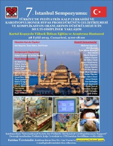
7th Istanbul Symposium of International Society for Pediatric Mechanical Cardiopulmonary Support

NAFEMS European Conference on Multiphysics Simulation 2018

11th & 12th October 2018
Budapest, Hungary

Technology is changing faster than ever. Global megatrends – such as digitalization, resource scarcity, and the need for renewable energy – drive the demand for innovation and efficient product development. In today’s world of almost limitless computing power, numerical simulations need to be both accessible and accurate in order to enable innovation.

NAFEMS are pleased to announce the fourth European Conference on Multiphysics simulations in October 2018. It will cover the use of Multiphysics simulations in industry.

Source

Functional blood vessels regenerated in vivo from human induced pluripotent stem cells

Vasculogenesis ? the process of blood vessel formation through a de novo production of endothelial cells (ECs, or those forming a thin layer lining the interior surface of blood and lymphatic vessels) ? is a vital tool in regenerative medicine, tissue engineering, and, in particular, the battle against vascular disease, the leading cause of mortality in the United States. (More than one in three Americans (36.9%) suffer from heart disease, and by 2030, an estimated 116 million people in the United States (40.5%) will have some form of cardiovascular disease.) More specifically, generating functional, long-lasting vasculogenic cells is a key but elusive component in human induced pluripotent stem (hiPS) research. Recently, however, researchers at Harvard University and Massachusetts General Hospital successfully generated endothelial cells from healthy donors’ hiPS cells to form stable functional blood vessels in vivo. Moreover, they developed an approach to generate mesenchymal precursor cells (MPCs, or multipotent stromal, or connective tissue, cells that can differentiate into a variety of cell types including perivascular cells ? another component of vessel wall) from hiPS cells in parallel, and also generated functional blood vessels in vivo using these endothelial and multipotent stromal derived cells from the same hiPS cell line. Beyond this, and in terms of clinical translation, the team successfully generated ECs and MPCs from Type 1 Diabetic patient-derived hiPS cell lines and also used them to generate blood vessels in vivo.

Read more

6.Istanbul Symposium: The Progresses and Conclusions on The Life Support Systems in Turkey /2013

The International Society for Pediatric Mechanical Cardiopulmonary Support organized 6.Istanbul Symposium: The Proggresses and Conclucions on The Life support Systems in Turkey /2013 Dr. Siyami Ersek Chest & Cardiovascular Surgery Training & Research Hospital.

Click for comitee members, speakers and panel topics.

All partipiants had a sertificate and the english translation of Aydın Aytaç’s book named Life Dedicated to The Heart.

I specially thank Atıf Akçevin for his advises and supports to my research topic and of course Kerem Pekkan and Ahmet Şaşmezel for their kind favors.

Stents disrupt blood flow

A researcher at ETH Zurich is designing a realistic artery model with an implanted stent and is using a computer to simulate the blood flow through the stent. In doing so he is uncovering weaknesses in this common form of therapy for atherosclerosis and paving the way for the development of optimized stents.

Coronary arteries are susceptible to plaque that can lead to illness, a phenomenon known informally as a “hardening” of the arteries. The effect of this is narrowing of blood vessels. If a clot detaches itself in another blood vessel and does not flow through the constricted section, this prevents blood supply to the heart and can be dangerous or even fatal in some cases. The disease-induced narrowing of coronary vessels and related heart attacks are one of the most common causes of death in industrial nations. Cardiologists use what are referred to as stents to open the closed-up vessels. Stents are tiny mesh tubes that are inserted into the bloodstream via the artery in the groin and placed in the constricted section. The doctors use a balloon catheter to expand the tube. This expands the blood vessel and the blood can flow through freely again. This method has saved the lives of many heart patients, but it does also have one disadvantage: in two of five patients, plaque and growths ? referred to as “restenosis” ? accumulate again at the place where the stent is located in the blood vessel, leading to a narrowing of the arteries once again. Arteries on the screen In order to understand the processes and the effects of stents in arteries better, Farhad Rikhtegar, a doctoral student from Professor Dimos Poulikakos’ group at ETH Zurich has implanted a stent in a porcine coronary artery in a laboratory, rendered the real internal surface of the artery including the stent in visual form and in mathematical terms and reproduced it exactly. He also created a computer model of the blood flow in the region of the stent. The paper relating to this work was recently published in the journal PloS One.

Rikhtegar’s computer models are realistic and achieve a level of detail that has never been seen before. Older models depicted blood vessels in a simplified manner and took into account only very simple clinical scenarios. However, the simulations by the ETH Zurich researcher are more complex and are based on real-life materials: porcine coronary arteries scanned at high resolution in which stents were implanted. Stents eliminate shear stress The simulations make clear that the tiny mesh-like strut connectors of a stent that lie against the wall of the blood vessel slow down the blood flow along these struts considerably. They also show that the blood in the tunnels formed by the stent struts can be “trapped” in vortices, which also reduces the speed of the blood flow in the vicinity of the stent. Because of the slower blood flow, the shear stress that the blood flow exerts on the wall of the blood vessel decreases. Endothelial cells that cover the blood vessel can only carry out their function properly if the blood flow and thus the shear stress acting on them reach a certain average value. If this stress is too low, they allow more and more bad cholesterol into the vessel wall. This in turn leads to infections that result in atherosclerosis once again. It can happen that the artery narrows once again in the region of the stent. Blind spots create a higher risk of restenosis The simulations also show what can happen if a stent is poorly positioned and does not lie flat against the vessel wall. In such a case, “blind spots” occur between the stent and the blood vessel wall where the speed of the blood flow is also severely reduced, increasing the risk of restenosis. Rikhtegar created further models where two stents, partially overlapping, were inserted into porcine arteries. The risk of restenosis is even greater when stents overlap than in the case of one individual stent. Evaluation of this data is not yet complete, however, and the doctoral student would like to publish the results in another paper. Intricate methodology Farhad Rikhtegar had to be quite creative with ideas on how to create the experiments and the related simulations. He created real-life conditions for the subsequent digitalization process using porcine hearts from the slaughterhouse. In collaboration with cardiologists from the University Hospital Zurich, he implanted the stents into the largest coronary artery. The ETH Zurich doctoral student then prepared the blood vessels by injecting them with resin. After the resin had hardened, he removed the tissue and thus obtained a three-dimensional sculpture of the blood vessels in the porcine heart. This preparation method is not new. However, Rikhtegar optimized the method by developing a pump that pressed the resin into the veins using physiological pressure. He explained that he was still injecting the resin by hand in the first trials. But he exerted too much pressure when using this method, destroying the fine capillaries in the vessels. By contrast, automated injection of the resin into the heart vessels ensured that the detailed meshing of even the finest capillaries was retained. Rikhtegar scanned this resin cast at high resolution with a micro-computed tomographer (CT) from Professor Ralph Müller’s group. This gave him a precise digital presentation of the artery in the vicinity of the stent, through which he was able to allow blood to flow in computer simulations. Normal computed tomography used in clinics and hospitals does not have a sufficient resolution to depict the struts of the stent, which are just 100 micrometres thick. However, micro-computed tomography can provide resolutions for structures of just a few micrometres in size. This allowed Rikhtegar to also present the deformation of the blood vessel wall in a vertical and horizontal direction and to show the position of unfavourably positioned stents. Using the geometry of the vein area containing the stent as calculated by the micro CT images, the ETH Zurich doctoral student was able to carry out simulations for almost any scenario. From aeronautics to medical applications Rikhtegar explains that the work was quite a challenge. There was a specific method for each individual sub-step. In cooperation with the respective experts in the field, the aim was to optimize each of these sub-steps. “That took a lot of time”, says Rikhtegar, who is originally an aeronautical engineer. For his dissertation, he had to acquire specific medical knowledge and learn the corresponding methods. “Now I can even insert stents into coronary arteries ? it’s not really that difficult”, he grins. In order to carry out his work, he used around 80 porcine hearts that he was able to get from the slaughterhouse. He used these to test and optimize the different work steps. He then had 15 hearts for the scans and the simulations. The method he has developed is a unique tool that allows for research into the relationship between hemodynamic factors and vascular biology. But the simulation can also be used to optimize the use of stents or to develop new forms of stents and test these on a computer. More information: Rikhtegar F, Pacheco F, Wyss C, Stok KS, Ge H, et al. (2013) Compound Ex Vivo and In Silico Method for Hemodynamic Analysis of Stented Arteries. PLoS ONE 8(3): e58147. doi:10.1371/journal.pone.0058147

Read more.

Air pollution and hardening of arteries

The researchers, led by Sara Adar, John Searle Assistant Professor of Epidemiology, University of Michigan School of Public Health, and Joel Kaufman, Professor of Environmental and Occupational Health Sciences and Medicine, University of Washington, found that higher concentrations of fine particulate air pollution (PM2.5) were linked to a faster thickening of the inner two layers of the common carotid artery, an important blood vessel that provides blood to the head, neck, and brain. They also found that reductions of fine particulate air pollution over time were linked to slower progression of the blood vessel thickness. The thickness of this blood vessel is an indicator of how much atherosclerosis is present in the arteries throughout the body, even among people with no obvious symptoms of heart disease. “Our findings help us to understand how it is that exposures to air pollution may cause the increases in heart attacks and strokes observed by other studies,” Adar said.

Read more

5th ISTANBUL SYMPOSIUM: BIOENGINEERING APPROACHES ON PEDIATRIC CARDIOVASCULAR MEDICINE

Scientific Co-Chairs are Kerem Pekkan, PhD & Akif Ündar, PhD

Invitation to Attend

The 5th Istanbul Symposium is intended for medical and engineering students, nurses,
scientists, pediatric heart surgeons, engineers, cardiologists, intensivists, neonatologists,
anesthesiologists, neurologists, pediatric perfusionists, respiratory therapists, residents
and fellows. All are invited and encouraged to attend.

Koç University, Engineering Auditorium ( Mühendislik Oditoryumu ) / 19 April 2013

International Congress of Cardiovascular Technologies /Algarve

Extended Abstract Submission and Complete Paper Submission: April 24, 2013
Authors Notification: June 17, 2013
Camera Ready and Registration: July 8, 2013

Read More

International Symposium on Endovascular Therapeutics / Barcelona

International Symposium on Endovascular Therapeutics is a scientific event created in Barcelona in 2000.  Its main objective is to update Endovascular professionals on the most relevant innovations interacting with international and multispecialty faculty. The meeting is held biyearly in Barcelona.

PROGRAM CONTENTS
  • Lower limb revascularization evidences
  • Wound healing adjuvants
  • Pharmaceutical adjuvants
  • Carotid stenting update
  • Upper limb revascularization
  • Visceral arterial management update
  • Venous disorders
  • Vascular access issues
  • Innovative therapeutic and diagnostic technology: The VIVA perspective
  • AAA management concerns
  • TEVAR management concerns
  • Ascending aorta and arch  endovascular approaches
  • Heart interventionalism hot evidence
  • Neuro-intervention innovation
  • Global Endovascular Economic analysis
  • The best from LINC:  a selection of the most educational live cases
The most important features of this event are:
? Hands on opportunities
? Abstracts and challenging case competition presentations
? Vascular, cardiac and neurovascular topics
? Hot, cutting edge discussion topics

Wireless device powers implanted blood-pressure sensor, eliminating batteries

Researchers at A*STAR Institute of Microelectronics in Singapore are developing a prototype wireless device that powers an implanted blood-pressure sensor, eliminating the need to recharge or replace a battery.

The microscale electronic sensor monitors blood flow through artificial blood vessels. Surgeons use these prosthetic grafts to bypass diseased or clogged blood vessels in patients experiencing restricted blood supply, for example.

Over time, however, the graft can also become blocked. To avoid complete failure, blood flow through the graft must be monitored regularly, but existing techniques are slow and costly.

Monitoring blood flow rate inside prosthetic vascular grafts enables early detection of graft degradation and prevention of graft failure.

The implant is powered by a handheld external reader, which uses inductive coupling to wirelessly transfer energy. The team developed an ultralow-power application-specific integrated circuit (ASIC) for the implant designed for low-power (21.6 ?W) use.

The sensors are based on piezoresistive silicon nanowires. As blood flows over the sensor, the associated mechanical stresses induce a measurable increase in electrical resistance, proportional to the flow pressure.

?Our flow sensor system achieves an ultra-low power consumption of 12.6 microwatts,? said A*STAR?s Jia Hao Cheong, who heads the project. To achieve that the sensor transmits its data to the handheld reader passively, by backscattering some of the incoming energy. ?We have tested our system with 50-millimeter-thick tissue between the external coil and implantable coil, and it successfully extracted the pressure data from the implantable device.?

?The next step of the project is to integrate the system and embed it inside a graft for an experimental animal,? Cheong said.

Source: http://www.kurzweilai.net

Heart repair breakthroughs replace surgeon’s knife

Heart care is in the midst of a transformation. Many problems that once required sawing through the breastbone and opening up the chest for open heart surgery now can be treated with a nip, twist or patch through a tube.
These minimal procedures used to be done just to unclog arteries and correct less common heart rhythm problems. Now some patients are getting such repairs for valves, irregular heartbeats, holes in the heart and other defects?without major surgery. Doctors even are testing ways to treat high blood pressure with some of these new approaches.

All rely on catheters?hollow tubes that let doctors burn away and reshape heart tissue or correct defects through small holes into blood vessels.

“This is the replacement for the surgeon’s knife. Instead of opening the chest, we’re able to put catheters in through the leg, sometimes through the arm,” said Dr. Spencer King of St. Joseph’s Heart and Vascular Institute in Atlanta. He is former president of the American College of Cardiology. Its conference earlier this month featured research on these novel devices.

“Many patients after having this kind of procedure in a day or two can go home” rather than staying in the hospital while a big wound heals, he said. It may lead to cheaper treatment, although the initial cost of the novel devices often offsets the savings from shorter hospital stays.

Not everyone can have catheter treatment, and some promising devices have hit snags in testing. Others on the market now are so new that it will take several years to see if their results last as long as the benefits from surgery do.

But already, these procedures have allowed many people too old or frail for an operation to get help for problems that otherwise would likely kill them.

“You can do these on 90-year-old patients,” King said.

These methods also offer an option for people who cannot tolerate long-term use of blood thinners or other drugs to manage their conditions, or who don’t get enough help from these medicines and are getting worse. “It’s opened up a whole new field,” said Dr. Hadley Wilson, cardiology chief at Carolinas HealthCare System in Charlotte. “We can hopefully treat more patients more definitively, with better results.” For patients, this is crucial: Make sure you are evaluated by a “heart team” that includes a surgeon as well as other specialists who do less invasive treatments. Many patients now get whatever treatment is offered by whatever specialist they are sent to, and those specialists sometimes are rivals. “We want to get away from that” and do whatever is best for the patient, said Dr. Timothy Gardner, a surgeon at Christiana Care Health System in Newark, Delaware, and an American Heart Association spokesman. “There shouldn’t be a rivalry in the field.” Here are some common problems and newer treatments for them:

Heart valves

illions of people have leaky heart valves. Each year, more than 100,000 people in the United States alone have surgery for them. A common one is the aortic valve, the heart’s main gate. It can stiffen and narrow, making the heart strain to push blood through it. Without a valve replacement operation, half of these patients die within two years, yet many are too weak to have one. “Essentially, this was a death sentence,” said Dr. John Harold, a Los Angeles heart specialist who is president of the College of Cardiology. That changed just over a year ago, when Edwards Lifesciences Corp. won approval to sell an artificial aortic valve flexible and small enough to fit into a catheter and be wedged inside the bad one. At first it was just for inoperable patients. Last fall, use was expanded to include people able to have surgery but at high risk of complications. Gary Verwer, 76, of Napa, California, had a bypass operation in 1988 that made surgery too risky when he later developed trouble with his aortic valve. “It was getting worse every day. I couldn’t walk from my bed to my bathroom without having to sit down and rest,” he said. After getting a new valve through a catheter last April at Stanford University, “everything changed; it was almost immediate,” he said. “Now I can walk almost three miles a day and enjoy it. I’m not tired at all.” “The chest cracking part is not the most fun,” he said of his earlier bypass surgery. “It was a great relief not to have to go through that recovery again.” Catheter-based treatments for other valves also are in testing. One for the mitral valve?Abbott Laboratories’ MitraClip?had a mixed review by federal Food and Drug Administration advisers this week; whether it will win FDA approval is unclear. It is already sold in Europe.

Heart rhythm problems

Catheters can contain tools to vaporize or “ablate” bits of heart tissue that cause abnormal signals that control the heartbeat. This used to be done only for some serious or relatively rare problems, or surgically if a patient was having an operation for another heart issue. Now catheter ablation is being used for the most common rhythm problem?atrial fibrillation, which plagues about 3 million Americans and 15 million people worldwide. The upper chambers of the heart quiver or beat too fast or too slow. That lets blood pool in a small pouch off one of these chambers. Clots can form in the pouch and travel to the brain, causing a stroke. Ablation addresses the underlying rhythm problem. To address the stroke risk from pooled blood, several novel devices aim to plug or seal off the pouch. Only one has approval in the U.S. now?SentreHeart Inc.’s Lariat, a tiny lasso to cinch the pouch shut. It uses two catheters that act like chopsticks. One goes through a blood vessel and into the pouch to help guide placement of the device, which is contained in a second catheter poked under the ribs to the outside of the heart. A loop is released to circle the top of the pouch where it meets the heart, sealing off the pouch. A different kind of device?Boston Scientific Corp.’s Watchman?is sold in Europe and parts of Asia, but is pending before the FDA in the U.S. It’s like a tiny umbrella pushed through a vein and then opened inside the heart to plug the troublesome pouch. Early results from a pivotal study released by the company suggested it would miss a key goal, making its future in the U.S. uncertain.

Heart defects

Some people have a hole in a heart wall called an atrial septal defect that causes abnormal blood flow. St. Jude Medical Inc.’s Amplatzer is a fabric-mesh patch threaded through catheters to plug the hole. The patch is also being tested for a more common defect?PFO, a hole that results when the heart wall doesn’t seal the way it should after birth. This can raise the risk of stroke. In two new studies, the device did not meet the main goal of lowering the risk of repeat strokes in people who had already suffered one, but some doctors were encouraged by other results.

?logged arteries

The original catheter-based treatment?balloon angioplasty?is still used hundreds of thousands of times each year in the U.S. alone. A Japanese company, Terumo Corp., is one of the leaders of a new way to do it that is easier on patients?through a catheter in the arm rather than the groin. Newer stents that prop arteries open and then dissolve over time, aimed at reducing the risk of blood clots, also are in late-stage testing.

High blood pressure

About 75 million Americans and 1 billion people worldwide have high blood pressure, a major risk factor for heart attacks. Researchers are testing a possible long-term fix for dangerously high pressure that can’t be controlled with multiple medications. It uses a catheter and radio waves to zap nerves, located near the kidneys, which fuel high blood pressure. At least one device is approved in Europe and several companies are testing devices in the United States. “We’re very excited about this,” said Harold, the cardiology college’s president. It offers hope to “essentially cure high blood pressure.” Copyright 2013 The Associated Press. All rights reserved. This material may not be published, broadcast, rewritten or redistributed.

Read more at: http://medicalxpress.com/news/2013-03-heart-breakthroughs-surgeon-knife.html#jCp


CT scans find clogged arteries in mummies

“We found that heart disease is a serial killer that has been stalking mankind for thousands of years,” says researcher Gregory Thomas. “In the last century, atherosclerotic vascular disease has replaced infectious disease as the leading cause of death across the developed world.”

The researchers performed CT scans of 137 mummies from across four continents and found artery plaque in every single population studied, from preagricultural hunter-gatherers in the Aleutian Islands to the ancient Puebloans of southwestern United States.

Their findings provide an important twist to one?s understanding of atherosclerotic vascular disease, which is the leading cause of death in the developed world: While modern lifestyles can accelerate the development of plaque on arteries, the prevalence of the disease across human history shows it may have a more basic connection to inflammation and aging.

?This is not a disease only of modern circumstance but a basic feature of human aging in all populations,? says Professor Caleb Finch of University of Southern California (USC), a senior author of the study.

?Turns out even a Bronze Age guy from 5,000 years ago had calcified, carotid arteries,? Finch says, referring to Ötzi the Iceman, a natural mummy who lived around 3200 B.C. and was discovered frozen in a glacier in the Italian Alps in 1991.

With Gregory Thomas of Long Beach Memorial, Finch was part of a team that previously showed Egyptian mummies had calcified patches on their arteries indicative of advanced atherosclerosis (from the Greek ?athero,? meaning ?gruel,? and ?scler,? meaning ?hard?).

But ancient Egyptians tended to mummify only royalty or those who had privileged lives. The new study led by Thomas and Randall Thompson of Saint Luke?s Mid America Heart Institute examined mummies from four drastically different climates and diets?and from cultures that mummified regular people, including ancient Peruvians, Ancestral Puebloans, the Unangans of the Aleutian Islands, and ancient Egyptians.

?Our research shows that we are all at risk for atherosclerosis, the disease that causes heart attacks and strokes?all races, diets, and lifestyles,? says Thomas.

?Because of this we all need to be cautious of our diet, weight, and exercise to minimize its impact. The data gathered about individuals from the prehistoric cultures of ancient Peru and the Native Americans living along the Colorado River and the Unangan of the Aleutian Islands is forcing us to think outside the box and look for other factors that may cause heart disease.?

Overall, the researchers found probable or definite atherosclerosis in 34 percent of the mummies studied, with calcification of arteries more pronounced in the mummies that were older at the time of death. Atherosclerosis was equally common in mummies identified as male or female.

?We found that heart disease is a serial killer that has been stalking mankind for thousands of years,? Thompson says. ?In the last century, atherosclerotic vascular disease has replaced infectious disease as the leading cause of death across the developed world.

?A common assumption is that the rise in levels of atherosclerosis is predominantly lifestyle-related, and that if modern humans could emulate preindustrial or even preagricultural lifestyles, that atherosclerosis, or at least its clinical manifestations, would be avoided.

?Our findings seem to cast doubt on that assumption, and at the very least, we think they suggest that our understanding of the causes of atherosclerosis is incomplete and that it might be somehow inherent to the process of human aging,? he adds.

The researchers will next seek to biopsy ancient mummies to get a better understanding of the role chronic infection, inflammation, and genetics play in promoting the prevalence of atherosclerosis.

?Atherosclerosis starts very early in life. In the United States, most kids have little bumps on their arteries. Even stillbirths have little tiny nests of inflammatory cells. But environmental factors can accelerate this process,? Finch says, pointing to studies that show larger plaque buildup in children exposed to household tobacco smoking or who are obese.

Source: University of Southern California

Infant brain controls blood flow differently

?The control of blood flow in the brain is very important,? says Elizabeth Hillman, associate professor of biomedical engineering and of radiology, who led the research study in her Laboratory for Functional Optical Imaging at Columbia University.

?Not only are regionally specific increases in blood flow necessary for normal brain function, but these blood-flow increases form the basis of signals measured in fMRI, a critical imaging tool used widely in adults and children to assess brain function,? says Hillman. ?Many prior fMRI studies have overlooked the possibility that the infant brain controls blood flow differently.?

Functional magnetic resonance imaging, or fMRI, is one of several brain-imaging methods that measure changes in blood flow to detect the presence and location of neuronal activity. In adults, blood-flow increases occur in specific regions of the brain during a particular task like moving your hand or reacting to a stimulus.

?We found that the immature brain does not generate localized blood-flow increases in response to stimuli,? says Mariel Kozberg, a neurobiology MD-PhD candidate and lead author of the paper published in the Proceedings of the National Academy of Sciences. ?By tracking changes in blood-flow control with increasing age, we observed the brain gradually developing its ability to increase local blood flow and, by adulthood, generate a large blood-flow response.?

Hillman says the findings suggest that vascular development may be an important new factor to consider in normal and abnormal brain development.

The team used a unique high-speed, high-resolution imaging approach that takes advantage of the different absorption spectra of deoxygenated and oxygenated hemoglobin in order to determine changes in the concentrations of each.

The researchers found that, with increasing age, there was a gradual development of a localized increase in blood flow, while a strong, delayed decrease in flow was consistently present. Only by adulthood was the positive increase able to balance the decrease in flow.

?Our results suggest that the infant brain might not be able to generate localized blood-flow increases, even if there is neuronal activity occurring, and that the development of blood-flow control occurs in parallel with early neuronal development,? says Kozberg.

?This could suggest that fMRI studies of infants and children may be detecting changes in both vascular and neuronal development?in fact, vascular development may be an important new factor to consider in normal and abnormal brain development.?

The team also found that the younger age groups were highly sensitive to blood pressure increases in response to stimulation and that these increases can cause large increases in blood flow across the brain.

?This finding indicates that the newborn brain is also unable to regulate its overall blood-flow levels,? Kozberg explains. ?This could explain earlier fMRI results in infants and children that were sometimes positive and sometimes negative, because it is difficult to tell whether blood pressure increases are occurring in infants and children. This result suggests that great care should be taken in setting stimulus thresholds in young subjects.?

The researchers add that, since the newborn brain appears to be able to sustain itself without tightly controlled blood flow, their findings suggest that the infant brain may be intrinsically more resistant to damage due to a lack of oxygen than the adult brain.

?This could be an important property to understand, both in terms of understanding how best to treat blood-flow problems in the newborn infant brain, which can cause lifelong problems such as cerebral palsy, and to potentially better understand how to treat the adult brain in conditions such as stroke,? Hillman observes.

This research was supported by grants and student fellowships from the National Institute of Neurological Disorders and Stroke, the National Eye Institute, the National Science Foundation, the National Defense Science and Engineering Graduate Fellowship, the Medical Scientist Training Program, and the Human Frontier Science Program.

SourceColumbia University

Blood plasma

Blood plasma is placed between two plates and the plates then drawn apart. High-speed cameras fitted with high-resolution microscope lenses capture the formation of threads and drops, demonstrating that blood plasma exhibits both viscous and elastic behavior when deformed and that it does not behave like water. Credit: Christof Schäfer, Phys. Rev. Lett. 110, 2013, 078305. Copyright (2013) by the American Physical Society

Read more at: http://medicalxpress.com/news/2013-02-blood-thicker-waterand-plasma-video.html#jCp

Gender influences ischemic time, outcomes after STEMI

After ST-segment elevation myocardial infarction and primary percutaneous coronary intervention, women have longer ischemic times and are at a higher risk than men of early all-cause and cardiac mortality, according to research published in the Feb. 1 issue of The American Journal of Cardiology.

Read more at: http://medicalxpress.com/news/2013-02-gender-ischemic-outcomes-stemi.html#jCp

Video capsule accurately detects intestinal blood

Video capsule endoscopy can be safely and accurately used to detect blood in patients with acute upper gastrointestinal hemorrhage seen in emergency departments, according to a study published online Feb. 11 in the Annals of Emergency Medicine.

Read more at: http://medicalxpress.com/news/2013-02-video-capsule-accurately-intestinal-blood.html#jCp

Researchers show how blood vessels regroup after stroke

Growth factors released by oxygen-starved cells prompt nearby endothelial cells, which line blood vessels, to grow into new networks. Researchers at Rice University are working to understand how to direct the process in the brains of stroke and disease patients. Credit: Qutub Lab/Rice University Rice scientists simulate “robot” cells to study the development of microvascular systems in the brain. The goal is to find a way to direct the development of vessels that feed oxygen-starved cells in stroke and neurodegenerative disease patients.

Read more at: http://medicalxpress.com/news/2013-02-blood-vessels-regroup.html#jCp

Virtual heart sheds new light on heart defect

Researchers at the School of Physics and Astronomy used cutting edge technology to build an advanced computational model of an anatomically correct sheep’s heart. It was made by taking a series of very thin slices of the heart, imaging them in 2D and then using a computer programme to render them into a 3D model. The reconstruction includes details of the complex fibre structure of the tissue, and the segmentation of the upper chambers of the heart into known distinctive atrial regions. Single-cell models that take into account information about the electrical activity in different atrial parts of regions the heart were then incorporated into the model. The virtual heart was then used to investigate the condition atrial fibrillation (AF). Professor Henggui Zhang led the research and explains why they wanted to study AF: “Atrial fibrillation (AF) affects approximately 1.5% of the world’s population. In the UK more than 500,000 patients have been diagnosed with the condition which causes an irregular heart rate. It is also known to increase the risk and severity of stroke. Despite its prevalence very little is known about what causes AF. We hoped our model would allow us to understand the mechanisms of this condition to ultimately help create better treatments.” AF occurs when abnormal electrical impulses suddenly start firing in the upper chambers of the heart. These impulses override the heart’s natural pacemaker, which can no longer control the rhythm of the heart. This desynchronises the heart muscle contraction and reduces the heart’s efficiency and performance. Professor Zhang and his team focussed on the pulmonary vein which is a common area that initially triggers AF. They simulated erratic electrical waves passing through the vein and the surrounding atrial tissue, and then studied the impact this had on the rest of the heart. What they found was that regional differences in the electrical activity across the tissue of the heart, known as electrical heterogeneity, is key to the initiation of AF. The largest electrical difference was between the pulmonary vein and the left atrium which may go some way to explaining why the pulmonary vein region is a common source of irregular heartbeats. The scientists also identified that the fibre structure of the heart plays an important role in the development of AF. There were directional variations in the conduction of electrical waves along and across the fibres, this is known as anisotropy. The fibre structure in the left atrium is much more organised compared with the complex structures of the pulmonary vein region. The sudden variation in conduction at the junction between the left atrium and the pulmonary vein regions appeared to contribute to the development of AF. Professor Zhang says: “This study has for the first time identified the individual role of electrical heterogeneity and fibre structure in the initiation and development of AF. It has not previously been possible to study the contribution of the two separately but using our computational model we’ve been able to clearly see that both electrical heterogeneity and fibre structure need to be taken into consideration when treatment strategies for AF are being devised.” The next step for Professor Zhang and his team will be to find a way to target the electrical conduction in specific regions of the heart to better protect against AF. They also want to use their virtual heart to gain a deeper understands of AF and to apply their findings to the development of more effective treatments. Professor Zhang concludes: “We’re really excited about the potential that our virtual heart opens up for research into this incredibly complex organ. By bringing together physics and biology we hope to unlock some of the unanswered questions about atrial fibrillation ? a condition which is only going to become more common as people live longer.” More information: The paper “A novel computation sheep atria model for the study of atrial fibrillation” will be published online in the Royal Society’s independent journal, Interface Focus, on Wednesday 16 January 2013. Provided by University of Manchester

Read more at: http://medicalxpress.com/news/2013-01-virtual-heart-defect.html#jCp

Source : Medical Express

5th ISTANBUL SYMPOSIUM: BIOENGINEERING APPROACHES ON PEDIATRIC CARDIOVASCULAR MEDICINE

5th ISTANBUL SYMPOSIUM: BIOENGINEERING APPROACHES ON PEDIATRIC CARDIOVASCULAR MEDICINE – Koç University, Sevgi Gönül Auditorium – 21 December 2012

Scientific Co-Chairs are Kerem Pekkan, PhD & Akif Ündar, PhD

Invitation to Attend

The 5th Istanbul Symposium is intended for medical and engineering students, nurses,
scientists, pediatric heart surgeons, engineers, cardiologists, intensivists, neonatologists,
anesthesiologists, neurologists, pediatric perfusionists, respiratory therapists, residents
and fellows. All are invited and encouraged to attend.

Invited Faculty

Mehmet A. Ağırbaşlı, MD Dept. of Cardiology, Marmara University, Istanbul, Türkiye
Atıf Akçevin, MD Dept. of Cardiovascular Surgery, Medipol University, Istanbul, Türkiye
Tijen Alkan-Bozkaya, MD Dept. of Cardiovascular Surgery, Medipol University, Istanbul, Türkiye
Ihsan Bakır, MD Istanbul Mehmet Akif Ersoy Thoracic and Cardiovascular
Surgery Training and Research Hospital, Istanbul, Türkiye
Hakan Ceyran, MD Istanbul Koşuyolu Heart Hospital, Cardiovascular Surgery,
Istanbul, Türkiye
Sertaç Haydın, MD Istanbul Mehmet Akif Ersoy Thoracic and Cardiovascular
Surgery Training and Research Hospital, Istanbul, Türkiye
Ender Ödemiş, MD Istanbul Mehmet Akif Ersoy Thoracic and Cardiovascular
Surgery Training and Research Hospital, Istanbul, Türkiye
Kerem Pekkan, PhD Şevket Ruacan, MD, Dept. Of Mechanical Engineering, Koc University, Istanbul,
Türkiye
Dean, College of Medicine, Koç University, Istanbul, Türkiye
Ayda Türköz, MD Başkent University, Department of Anethesiology, Istanbul,
Türkiye
Rıza Türköz, MD Başkent University, Department of Cardiovascular Surgery,
Istanbul, Türkiye
Akif Ündar, PhD Penn State Hershey Pediatric Cardiovascular Research
Center, Penn State Hershey College of Medicine, Penn State
Hershey Children’s Hospital, Hershey, PA, USA
Songül Yaşar Yıldız, PhD Candidate Dept. of Bioengineeering, Marmara University,
Istanbul, Türkiye)
TBA (Koç University, School of Nursing, Istanbul, Türkiye

3
SCIENTIFIC PROGRAM
9:00 ? 9:20 am Welcome ? Kerem Pekkan, PhD& Şevket Ruacan, MD, Dean, College of Medicine, Koç University,
Istanbul, Türkiye
9:20 ? 10:00 am ABC’s of Pediatric Cardiovascular Research for Medical and
Engineering Students
Akif Ündar, PhD ? Penn State Hershey Pediatric Cardiovascular
Research Center, Penn State Hershey College of Medicine, Penn
State Hershey Children’s Hospital, Hershey, PA, USA
10:00 ? 11:00 am Key Note Lecture – Applications of Computational Fluid Dynamics to
solve pediatric cardiovascular problems
Kerem Pekkan, PhD, (Dept. Of Mechanical Engineering, Koc
University, Istanbul, Türkiye)
Introduction: Akif Ündar, PhD
11:00 ? 11: 15 am Break
11:15 am ? Noon CASE STUDY: Novel fenestration designs for controlled venous flow
shunting in failing Fontans with systemic hypertension
Cardiac Surgeon: Definition of the problem – Rıza Türköz, MD
(Başkent University, Department of Cardiovascular Surgery, Istanbul,
Türkiye)
Scientist: Suggested Solution – Kerem Pekkan, PhD
12:00 ? 13:30 pm Lunch
13:30 ? 14:00 pm What are the "real" problems for pediatric cardiac patients in
Türkiye? Why multi-disciplinary approach is necessity, not an option?
Atıf Akçevin, MD (Dept. of Cardiovascular Surgery, Medipol
University, Istanbul)
14:00 ? 15:30 pm PANEL: Importance of Multidisciplinary Team Approach to Improve
the Outcomes During and After Neonatal and Pediatric
Cardiopulmonary Bypass Procedures in Türkiye
Moderators: Atıf Akçevin, MD and Ihsan Bakır, MD

4
Pediatric Cardiologists’ Perspective – Ender Ödemiş, MD (Istanbul
Mehmet Akif Ersoy Thoracic and Cardiovascular Surgery Training and
Research Hospital, Istanbul, Türkiye) (20 min)
Pediatric Cardiac Surgeons’ Perspective – Hakan Ceyran, MD (Istanbul
Koşuyolu Heart Hospital, Cardiovascular Surgery, Istanbul, Türkiye)
(20 min)
Pediatric Anesthesiologists’ Perspective – Ayda Türköz, MD (Başkent University, Department of Anethesiology, Istanbul, Türkiye)
(20 min)
Pediatric Nurses’ Perspective – TBA (Koç University, School of Nursing, Istanbul, Türkiye (20 min)
15:30 ? 16:00pm Break
16:00 ? 18:00pm PANEL: Pediatric ECLS Systems & Novel Techniques and Methods to
Minimize the Injury during neonatal/Pediatric Cardiopulmonary
Bypass Procedures
Moderators: Hakan Ceyran, MD and Rıza Türköz, MD
Pediatric Extracorporeal Life Support Systems in Türkiye ? 2012
Update –
Sertaç Haydın, MD (Istanbul Mehmet Akif Ersoy Thoracic and
Cardiovascular Surgery Training and Research Hospital, Istanbul,
Türkiye)
Monitoring Biomarkers After Pediatric Cardiac Surgery: A New
Paradigm in the Horizon, Mehmet A. Ağırbaşlı, MD (Dept. of
Cardiology, Marmara University, Istanbul, Türkiye)
Extremophiles for cardiovascular research – Songül Yaşar Yıldız, PhD
Candidate (Dept. of Bioengineeering, Marmara University, Istanbul,
Türkiye)
Impact of Pulsatile Perfusion on Clinical Outcomes of Neonates and
infants with Complex Pathologies undergoing Cardiopulmonary
Bypass Procedures ?
Tijen Alkan-Bozkaya, MD (Dept. of Cardiovascular Surgery, Medipol
University, Istanbul)

5
Importance of Neonatal/Pediatric oxygenators with or without
Arterial Filters for capturing microemboli during CPB procedures –
Akif Ündar, PhD
18:00pm Closing Remarks – Kerem Pekkan, PhD

A Primer on Computational Simulation in Congenital Heart Disease for the Clinician

Interest in the application of engineering methods to problems in congenital heart disease has
gained increased popularity over the past decade. The use of computational simulation to
examine common clinical problems including single ventricle physiology and the associated
surgical approaches, the effects of pacemaker implantation on vascular occlusion, or
delineation of the biomechanical effects of implanted medical devices is now routinely
appearing in clinical journals within all pediatric cardiovascular subspecialties. In practice,
such collaboration can only work if both communities understand each other?s methods and
their limitations. This paper is intended to facilitate this communication by presenting in the
context of congenital heart disease (CHD) the main steps involved in performing
computational simulation – from the selection of an appropriate clinical question/problem to
understanding the computational results, and all of the ?black boxes? in between.

More

Handmade Glass Anatomical Models by Farlow’s Scientific Glassblowing

Gary Farlow can make art out of arteries. He and his team of 10 at Farlow?s Scientific Glassblowing are able to transform the body?s vasculature?and nearly all of its other parts?into an ornate borosilicate glass sculpture, from the heart?s ventricles to the brain?s circle of Willis. ?We do almost every part of the body,? Farlow says. ?It can take a pretty artistic mind to make some of these things.? With the help of cardiologists, the team creates custom see-through systems for science and medical training.

Their anatomically correct models can be designed to simulate blood flow, teach placement of catheters and angioplasty devices, or simply test or demo new surgical gizmos. Individual arteries, veins, and capillaries are shaped and fused together, one at a time. Ground-glass joints are added at the exposed ends so a head, say, can be connected to the carotid arteries should customers want to expand their model. A full-body setup could cost $25,000, so don?t get any bright ideas about using one as a brandy decanter.

4th Istanbul Symposium of Pediatric Life Support Systems and Pediatric Cardiopulmoner Bypass:The Last Advances in 2012

4th Istanbul Symposium of Pediatric Life Support Systems and Pediatric Cardiopulmoner Bypass:The Last Advances in 2012 hold on Istanbul Mehmet Akif Ersoy Thoracic & Cardiovascular Surgery Training and Research Hospital.

Atif AKCEVINSertac HAYDINEnder Odemis

Artifical Artery 1955

4th Istanbul Symposium: Pediatric Support Systems and Pediatric Cardiopulmonary Bypass

The 4th Pediatric Support Systems and Pediatric Cardiopulmonary

Bypass Symposium will be held on 28th July 2012

at Istanbul Mehmet Akif Ersoy

Thoracic & Cardiovascular Surgery Training and Research Hospital.

.

3th International VIB Ph.D. Symposium VIBes on Biosciences 2012

?The VIBes 2012 Organizing Committee has the pleasure to invite all Ph.D. Students to the 3rd edition of the VIB Ph.D. symposium ?VIBes in Biosciences 2012?, which will take place from 5 to 7 September 2012 in the city of Ghent (Belgium).

The VIBes 2012 program kicks off with several workshop sessions on the scientific publication process and soft skills training. The scientific program consists of two days of lectures by world-class scientists, covering a broad range of topics from immunology and molecular biology to cybernetics and biomedical engineering. Selected speakers will also discuss career opportunities and share insights on how to succeed in science.
VIBes 2012 will again be a very international event as we will provide up to 50 international (i.e. not working in Belgium) Ph.D. students with free attendance to the symposium, free accommodation as well as a limited amount of travel grants.

Registration is now open at www.vibes2012.org
Places are limited and provided on a first-come-first-serve basis.

The VIBes2012 Organizing Committee
Jonas Bethuyne, Miguel Lopez Cardoso, Eleonora Billi, Nathan De Geyter, Lynn Elton, Matthieu Moisse, Christina Müller, Julie Mutert, Zeynep Okray, Ioanna Petta, Sebastian Proost, Dariusz Ratman, Tim Snoek, Lorin Spruyt, Suresh Subedi, Joemar Taganna, Arun Kumar Tharkeshwar, Koen Tyberghein and Rajesh Vyas?

Cardiopulmonary Perfusion 8th International Conference on Pediatric Mechanical Circulatory Support Systems & Pediatric Cardiopulmonary Perfusion

Cardiopulmonary Perfusion 8th International Conference on Pediatric Mechanical Circulatory Support Systems & Pediatric Cardiopulmonary Perfusion will be held JUNE 13-16, 2012, GALATASARAY UNIVERSITY, ISTANBUL, TURKEY

Seminars 24th January

Biomedical Engineering Seminars’ speaker was  Kamuran KADIPAŞAOĞLU and this  has been the most incentive part of these meetings.

Image Source: Bahcesehir University

Frankfurt Euro Biotechnology Congress

Knee Anatomy Segmentation

I tried to show the knee anatomy with the MRI dataset of 3D Slicer (Harvard Medical School /Brigham and Women’s Hospital / Surgical Planning Laboratory).

Video: Knee Anatomy