Comments Off 
JUNE 4TH, 2021
By BANU PLUIE
A team I am honored to be involved in EVBio!
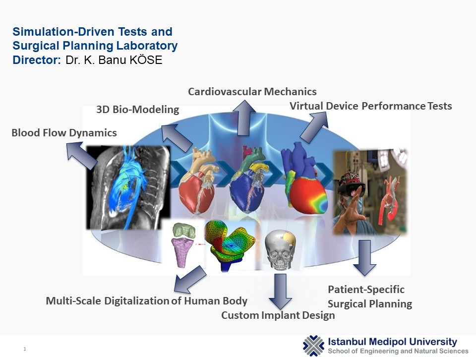
EVBio is a digital think tank formed by scientists from all disciplines related to vascular medicine, from molecular biology to scientific computing. Our mission is to imagine the future through disruptive basic and translational research. Our team works tirelessly to formulate one universal coherent theory for vascular disease and support all global efforts towards its conception and validation.

Our membership will increase soon!
Comments Off 
JANUARY 15TH, 2021
By BANU PLUIE
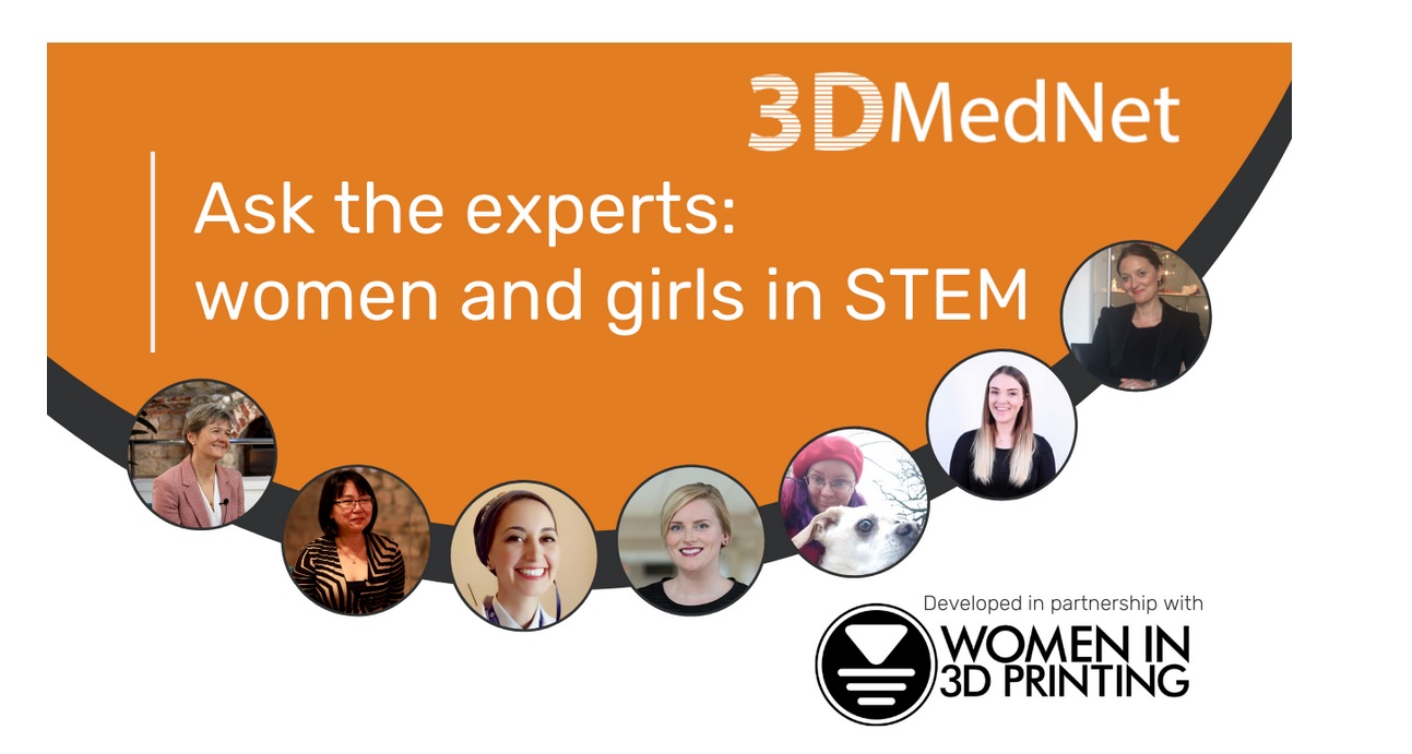
To commemorate International Women’s Day (8 March 2020), 3DMedNet has put together our first ‘Ask the experts’ feature in partnership with the global organization, Women in 3D Printing. Thanks to Georgi for inviting me to the conversation. Check the link for the full interview.
Comments Off 
FEBRUARY 21ST, 2020
By BANU PLUIE

To commemorate International Women’s Day on March 8, 2020, 3DMedNet will bring together our first ‘Ask the Experts’ feature in partnership with the global organization Women in 3D Printing! We invite you all to watch the live interview!
Stay Tuned! –> https://www.3dmednet.com/
Comments Off 
JULY 24TH, 2019
By BANU PLUIE
I was the guest of Women in 3D Printing this week.
The full-page is on this page: https://womenin3dprinting.com/banu-kose/
Thanks to Nora Toure for all the great work she has done and for bringing us together.
Read more »
Comments Off 
APRIL 16TH, 2018
By BANU PLUIE
The Mimics Innovation Suite (MIS) allows you to automate your workflows, potentially saving a lot of time, achieving more consistency, and reducing repetitive work and human error. That is an easy thing to say, but if you do not have much experience with scripting, we all know that it can be tough to get started. If you want to speed up your learning curve and get a head start, then this could be an interesting training for you.
Topics will include:
Basics of Optimizing your Workflow in Mimics 21 and 3-matic 13
How to write your first scripts
Introduction to Python
Hands-on training exercises for creating planning workflows (e.g. loading datasets, performing basic segmentation steps, landmarking, creating anatomical coordinate systems, designing custom implants)
Comments Off 
FEBRUARY 1ST, 2017
By BANU PLUIE
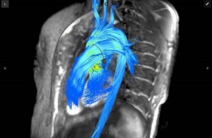
While cardiac magnetic resonance imaging (MRI) is considered an excellent imaging modality for the heart, offering highly detailed soft tissue anatomical imaging as well as functional assessments, it only makes up about 5 percent of all MRI scans in the United States. This is in part due to the expense, time involved and the complexity in completing these scans and reading them. There were two software innovations that may help increase the use of cardiac MRI by reducing its complexity.
To read the entire article, go to www.dicardiology.com/article/advances-cardiac-imaging-rsna-2016.
At RSNA 2015, Arterys introduced a package of advanced cardiac MRI visualization and quantification software that automates a lot of the processes involved. It also uses a cloud-based platform that allows access to a large amount of computing power needed to process cardiac cine functional data in real time. The software includes 4-D Flow and 2-D phase contrast workflows, and cardiac function measurements. The software is the first clinically available cardiovascular solution that delivers cloud-based, real-time processing of images with resolutions previously unattainable. The company gained U.S. Food and Drug Administration (FDA) 510(k) clearance in November 2016 and showed several new advancements at RSNA 2016. Arterys is partnering with GE Healthcare to introduce the software on the Signa MRI systems under the GE name of ViosWorks. However, Arterys said it has aspirations to be a software OEM for several MRI vendors. An additional introduction was Arterys? regurgitation evaluation software that offers several ways to view regurgitation, which has traditionally been difficult to assess on MRI. One view visualizes blood flow velocities with arrows to show direction of flow and a color code to show the speed of the flow. It presents very similar to cardiac ultrasound color flow Doppler. The software can help identify regurgitation jets, vortices and sheer wall stresses, and offers automated quantification. In cardiovascular research, sheer stress evaluation has become a big area of interest because it is believed these stresses may play a role in the formation of atherosclerosis, the degradation of heart valve function, and possibly play a role in the progression of heart failure. So, Arterys also introduced a research sheer stress analysis software package.
- DAVE FORNELL
To read the entire article, go to www.dicardiology.com/article/advances-cardiac-imaging-rsna-2016.
Comments Off 
JUNE 13TH, 2016
By BANU PLUIE
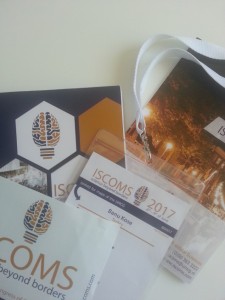
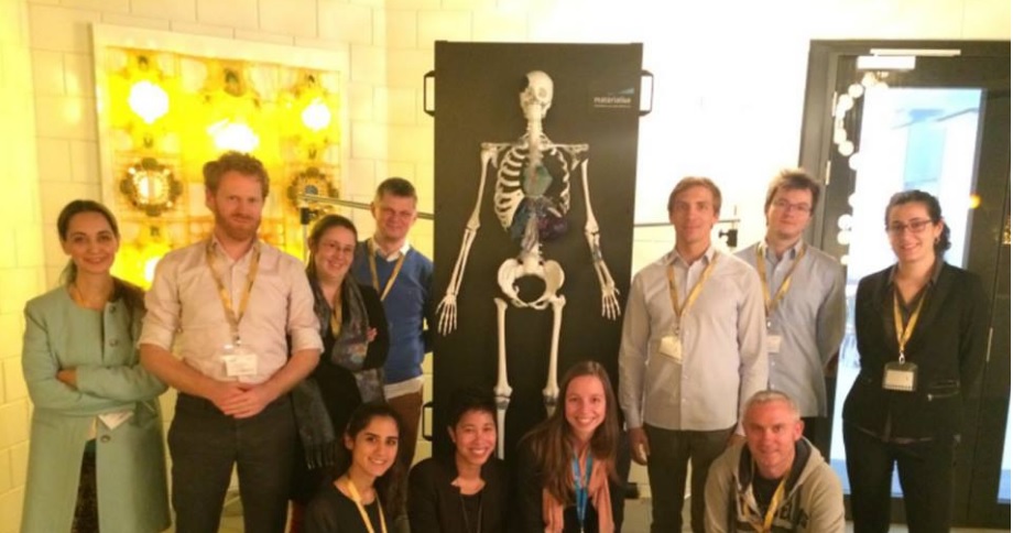
Many thanks to University Medical Center Groningen for the oral sessions and workshops of 3D Lab, LVAD treatment, Dissection of Brain, CABG treatment, IV Injections and Nuclear Medicine.

Comments Off 
MARCH 17TH, 2016
By BANU PLUIE

Materialise provided a Mimics Innovation Course on Soft Tissue.
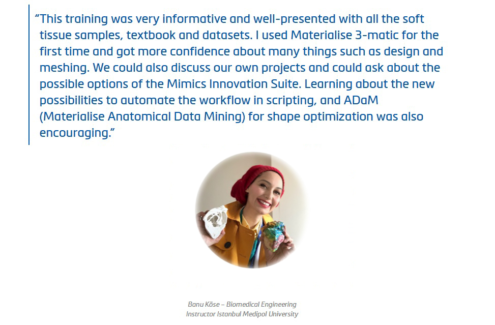
This training was very informative and well-presented with all soft tissue samples, text book and datasets.
I used 3-Matic for the first time, and got confidence about many things about design and meshing. We could also discuss our own projects and could ask possible options of Mimics Innovation Suite.
Learning about the news about scripting possibilities to automate the workflow, and ADam (Materialise Anatomical Data Mining) for shape optimisation was encouraging.
Thank you for sharing your knowledge with us, Karen de Leener and Inés da Silva.
Thank you for the great help of Job der Kinderen.
Comments Off 
JANUARY 2ND, 2016
By BANU PLUIE
In multidisciplinary areas, it is very important to be able to meet with the team who can work in harmony.
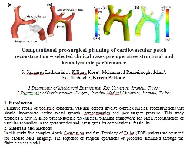
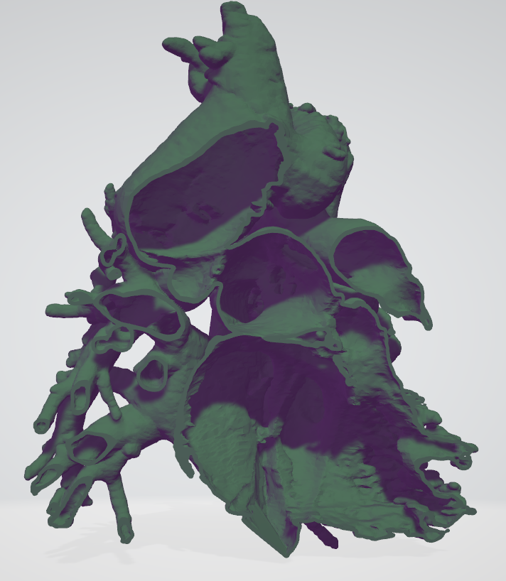
For his work on 3D intracardiac models on surgical planning, Thanks to cardiovascular surgeon Dr. Okan Yildiz, we have been informed about and contributed to many pediatric cases and treatments since 2016.
Comments Off 
NOVEMBER 9TH, 2015
By BANU PLUIE
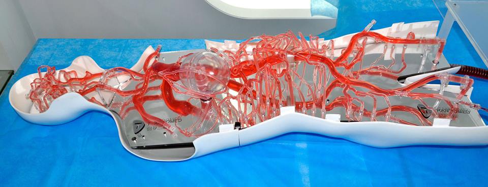
Trando Med will attend MEDICA 2017 in the Dusseldorf Germany from 13-16 November 2017. The booth is Hall 13 Booth F 9-05
Comments Off 
APRIL 15TH, 2015
By BANU PLUIE
Talking about cardiac imaging with Dr. Taliha Öner has always been a pleasure.
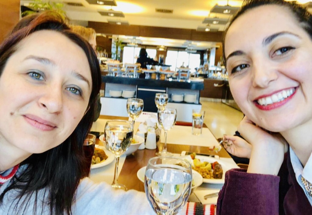
Good Luck in Boston.
Comments Off 
FEBRUARY 25TH, 2015
By BANU PLUIE
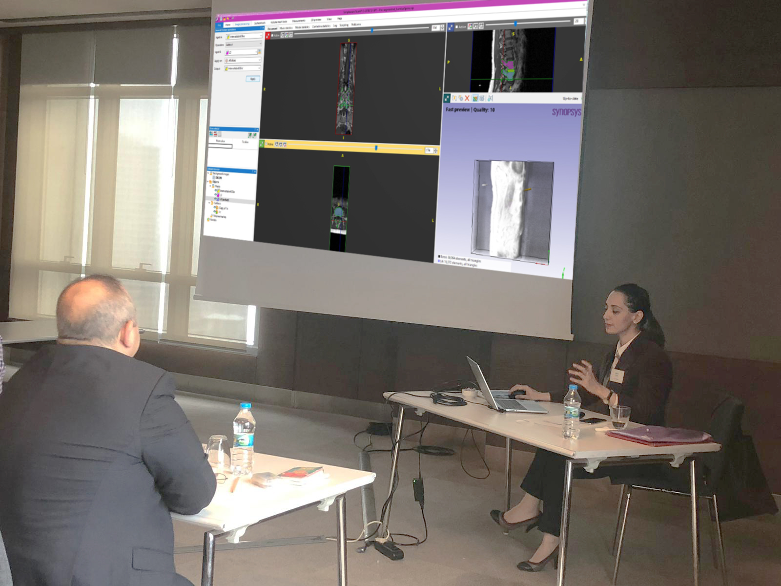
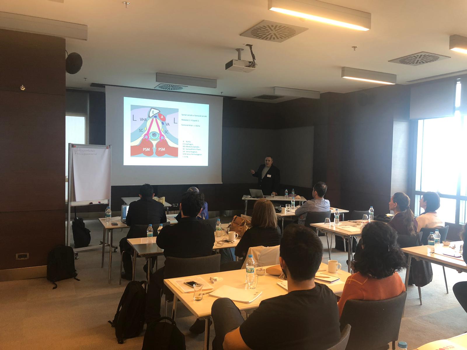
We depicted a live- surgical planning scenario with Prof. Erbil Oğuz and Kerem Girgin in Voksel 3D event. We used Simpleware for image processing, segmentation and designing.
Comments Off 
DECEMBER 31ST, 2014
By BANU PLUIE
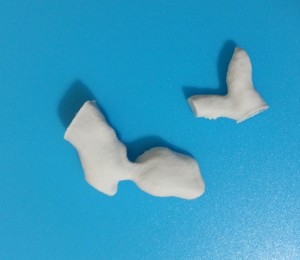
It was sensible showing 3D printed models when i was presenting my study.
Comments Off 
FEBRUARY 25TH, 2014
By BANU PLUIE
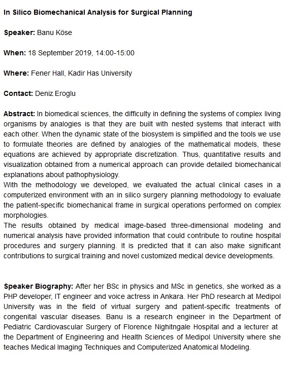
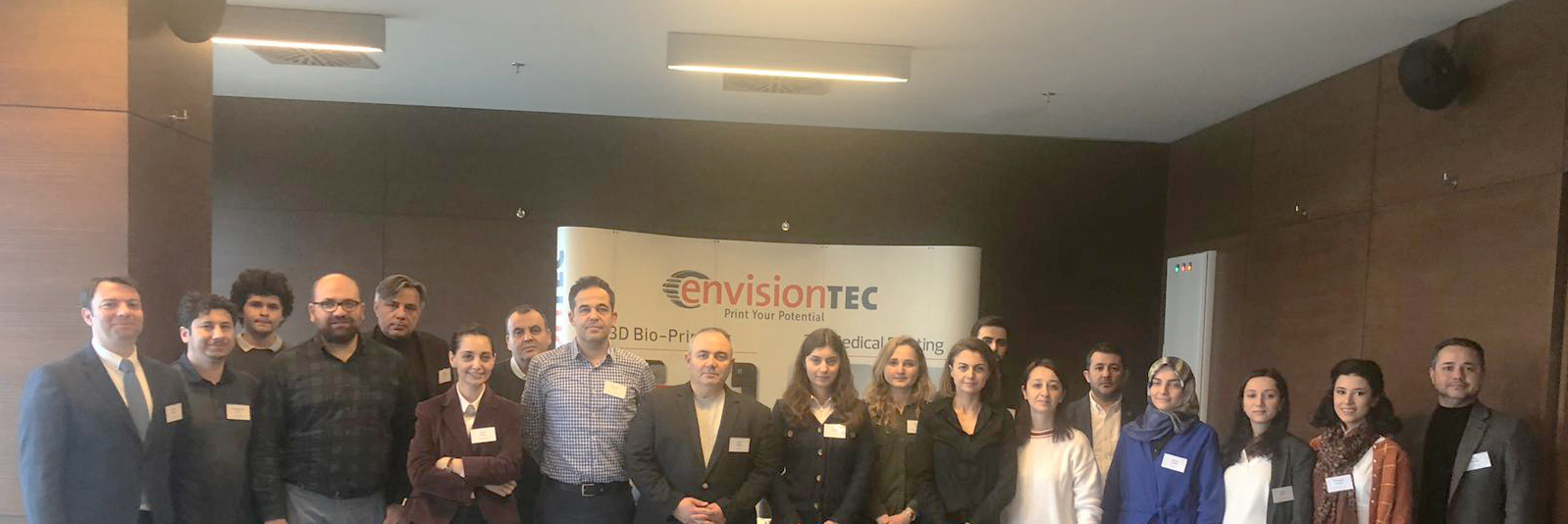
‘Voksel’s Anatomical Modeling, Surgical Planning, 3D Printing with Engineer – Surgeon Collaboration Training‘ was held on 23rd February in Istanbul.
I had the chance to share my experiences in image processing and modeling with the participants. I would like to thank Kerem Girgin, Erbil Oğuz, Samet Serbest and Cansu Çeltik from Voksel. It was great to be a part of Voksel team, and meeting with the participants who were aware of the benefits of interdisciplinary collaborations and patient-specific planning very well.
Comments Off 
NOVEMBER 5TH, 2013
By BANU PLUIE
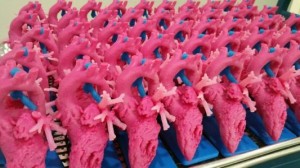
Alistair Phillips, MD, who is the Co-Chair for the American College of Cardiology, Surgeons Section tells about some of the impacts he has personally experienced using 3D printing in surgical settings as his participation in the 3DHEART program:
“The clinical trial is particularly exciting as it targets specific cases in which understanding of the anatomy will greatly enhance the surgical approach. A 3D printed replica of a patient’s heart will be created as part of the inclusion criteria to be in the study.Using 3D printing gave a better understanding of the Hybrid procedure, and allowed us to perform pulmonary valve replacement with a minimally invasive approach avoiding conventional method that required open-heart surgery. After coming to Cedars-Sinai we refined the pre-ventricular approach by utilizing a 3D printed models of patients’ hearts. We were able to simulate the implant into the right ventricular outflow tract.
Every surgeon is different. The education, experience, aptitudes, and attitude we bring to each equally nuanced and varied patient span an almost limitless spectrum and inform how we may utilize 3D printing for the benefit of our patients. The elegance of 3D printing is that it can create the individualized tools spanning this spectrum.
That said, however, what is not negotiable is the veracity of the models that we are receiving. Various materials and their corresponding colouring or rigidity may serve different functions in the hands of different surgeons, but ultimately we must have the utmost confidence in the fidelity of the models we are utilizing for pre-surgical planning. The more realistic the model is both in anatomical and textural preciousness will greatly enhance the application.
In all honesty, I would advise each hospital to start by really understanding the value proposition 3D printing offers across all specialities and, the culture of their institution. The best way to get answers to these very nebulous, complicated, nuanced directives is by retaining an outside vendor to provide as much of the services as possible, from the proverbial soup to nuts.
The excitement around the 3DHEART clinical trial is so great because it is the first organized, large-scale attempt to collect evidence of the efficacies of 3D printing in the practice of medicine and delivery of healthcare, not only in terms of optimized patient outcomes but also with respect to lower costs. If we can get reimbursement for 3D models, it is without a doubt a game-changer in terms of the practice of medicine, and a life-changer for many of our patients.”
Source
Comments Off 
NOVEMBER 5TH, 2013
By BANU PLUIE
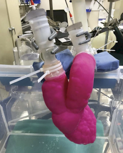
“Without the 3D printed models, we wouldn’t have been able to come up with a way to do the procedure in advance.”
—C. HUIE LIN, M.D
Adult congenital and interventional cardiologist.
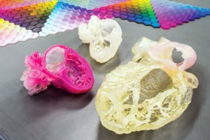
3D Print Bureau of Texas has partnered with physicians at Houston Methodist Hospital to create cardiac models for applications such as assessing the size and attachment site of a right atrial malignancy. Accurate physical replications of patient anatomy can even undergo testing in a dynamic system such as replicating the severity of aortic stenosis using flow testing.
3D Print Bureau of Texas also worked with Houston Methodist DeBakey Heart and Vascular Center on a complex case involving a young patient born with a wide-open leaking pulmonary valve. The patient could not take blood transfusions and have been turned down by two medical centres concerned she would not make it through surgery.
With a 3D printed model of the patient’s heart, Lin devised a plan that required very little blood loss, which resulted in a successful operation for the little patient.
Source
Comments Off 
JANUARY 15TH, 2010
By BANU PLUIE
I tried to show the knee anatomy with the MRI dataset of 3D Slicer (Harvard Medical School /Brigham and Women’s Hospital / Surgical Planning Laboratory).
Video: Knee Anatomy

![]() 3D Printing, 3D Slicer, ANSYS, Art, Artificial Organs, Banu Köse, Banu Pluie, BioFluidLab, Bioengineering, Biomaterials, Biomechanics, Biomed, Biomedical Science, Biophysics, Bioprinting, Biosecience, Cardiology, Cardiovascular Implants, Cerrahi, Circulatory Support Systems, ClinicianEngineerHub, Complex Systems, Congenital, Ece Salihoglu, Engineering, Finite Element Model, Fontan, Gradute, Heart, Heart Valves, Hemodynamics, Hepatic, IMAEH, Image Processing, K. Banu Köse, Kardiyovasküler, Kardiyovasküler Mekanik, Kevser Banu Köse, Laser Doppler, Liquid State, Materialise Medical, Medipol, Medtronic, Pediatric, Perfusion, Perfüzyon, PhD, Physics, Pulmonary, Radiology, Slicer, Structural Analysis, Surgery, SurgicalPlanning, Sıvı Hal Fiziği, TGA, The Journal of Cardiovascular Surgery, VSD, VirtalSurgery, Virtual Physiological Human, Windkessel, WomenEngineers, womenin3d
3D Printing, 3D Slicer, ANSYS, Art, Artificial Organs, Banu Köse, Banu Pluie, BioFluidLab, Bioengineering, Biomaterials, Biomechanics, Biomed, Biomedical Science, Biophysics, Bioprinting, Biosecience, Cardiology, Cardiovascular Implants, Cerrahi, Circulatory Support Systems, ClinicianEngineerHub, Complex Systems, Congenital, Ece Salihoglu, Engineering, Finite Element Model, Fontan, Gradute, Heart, Heart Valves, Hemodynamics, Hepatic, IMAEH, Image Processing, K. Banu Köse, Kardiyovasküler, Kardiyovasküler Mekanik, Kevser Banu Köse, Laser Doppler, Liquid State, Materialise Medical, Medipol, Medtronic, Pediatric, Perfusion, Perfüzyon, PhD, Physics, Pulmonary, Radiology, Slicer, Structural Analysis, Surgery, SurgicalPlanning, Sıvı Hal Fiziği, TGA, The Journal of Cardiovascular Surgery, VSD, VirtalSurgery, Virtual Physiological Human, Windkessel, WomenEngineers, womenin3d