Category: Art

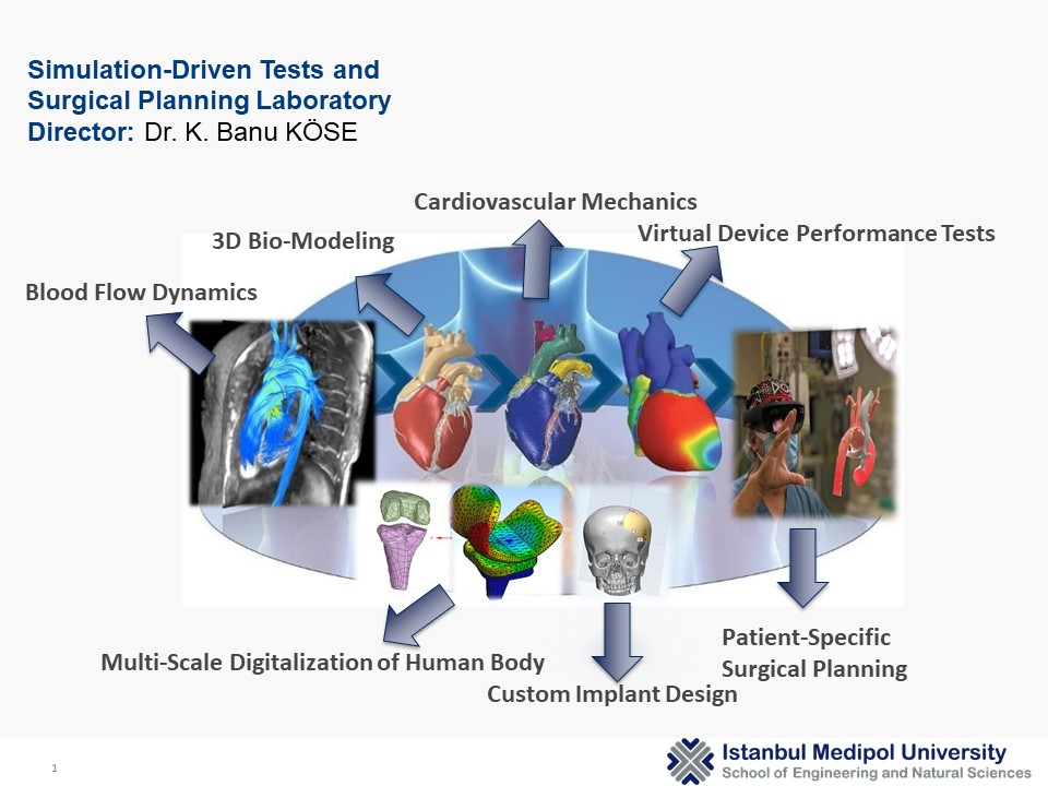
Surgical Planning Laboratory

MEDIPOL UNIVERSITY

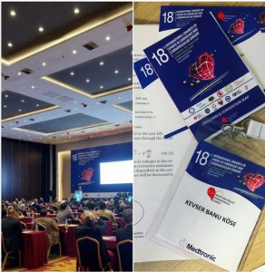
UCCVS 2022 Focus Valve Scholarship

I am very proud to have participated in 18th International Congress of Update Cardiology & Cardiovascular #Surgery with two research presentations, and ✨ to be awarded with FocusValve Scholarship
Best regards to #UCCVS committee members and our esteemed Prof. Öztekin Oto.

EVBio

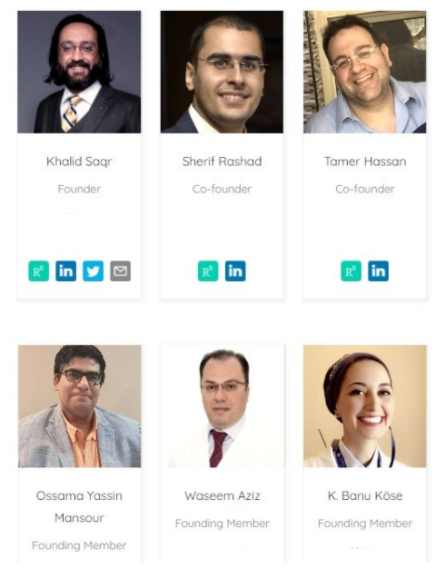
A team I am honored to be involved in EVBio!
EVBio is a digital think tank formed by scientists from all disciplines related to vascular medicine, from molecular biology to scientific computing. Our mission is to imagine the future through disruptive basic and translational research. Our team works tirelessly to formulate one universal coherent theory for vascular disease and support all global efforts towards its conception and validation.

Our membership will increase soon!

Liquid State Physics in Turkey

22. Liquid State Symposium (22. Sıvı Hal Senpozyumu) took place on 7th December 2018 in Piri Reis University.
It was very proud to be together with the physicist academics I knew and admired since my undergraduate years.
I find myself lucky to see the Prof. Zehra Akdeniz that I have always admired and exemplified. I could finally meet Prof. Nihat Berker who is not only a famous physicist but also an intellectual on comparative literature readings.

Thanks to Dr. Ozan Sarıyer and Dr. Gulsen Evingur for organizing this meeting.

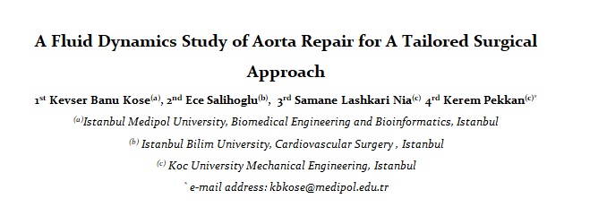
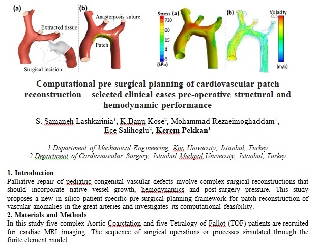
Prof Pekkan presented biological flow researches of his lab, and I presented a sample case of a pediatric aortic blood flow comparison study which is done with the great help of Dr. Ece Salihoglu.

Virtual Physiological Human Conference 2018 / Zaragoza

Conference Web Link

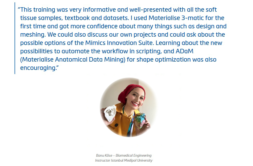
Optimizing your workflow in the Mimics Innovation Suite

The Mimics Innovation Suite (MIS) allows you to automate your workflows, potentially saving a lot of time, achieving more consistency, and reducing repetitive work and human error. That is an easy thing to say, but if you do not have much experience with scripting, we all know that it can be tough to get started. If you want to speed up your learning curve and get a head start, then this could be an interesting training for you.

Topics will include:

Basics of Optimizing your Workflow in Mimics 21 and 3-matic 13
How to write your first scripts
Introduction to Python
Hands-on training exercises for creating planning workflows (e.g. loading datasets, performing basic segmentation steps, landmarking, creating anatomical coordinate systems, designing custom implants)

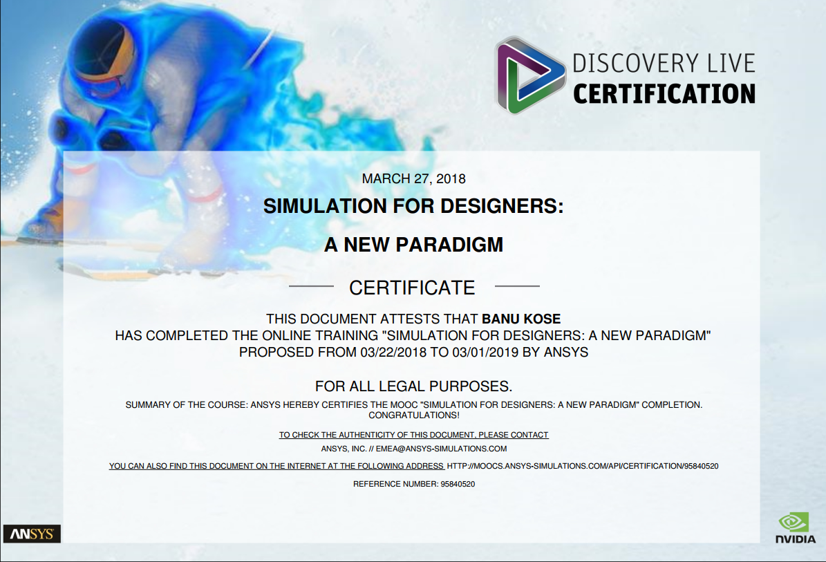
CERTIFICATED ANSYS DISCOVERY LIVE ’3D Modeling, Design & Simulation in real time’

HORST KIECHLE

Source

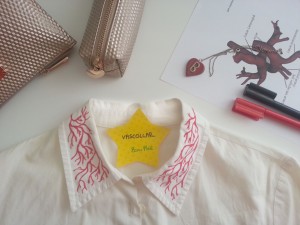
VasCollar (Vascular Collar)

I’ve just drown vessels to my collar. It looks better now.

For More  Drawords Stuff, Click Here.

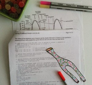
Windkessel plus colours ;)

I know why I’d rather CFD. Because it has colours. Windkessel analogy has not.

Link

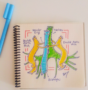
Double Aortic Arch

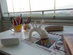
3D Printed Aorta

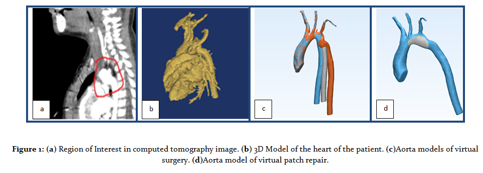
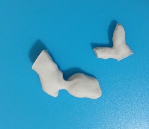
A pediatric aorta model reconstructed from the 3D CT images.

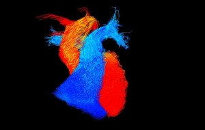
‘Go with the flow’ by Victoria Stoll

The British Heart Foundation (BHF) announced the winners of its annual ?Reflections of Research? image competition, reflecting the charity?s research into heart and circulatory disease.
The winning image ? titled ?Go with the flow,? by Victoria Stoll, a BHF-funded researcher at the University of Oxford ? captures the blood flowing within an adult heart frozen in time. Blood flows within the main pumping chambers (ventricles) of the heart and the vessels leaving the heart. The blue flow is blood that lacks oxygen and is travelling to the lungs. The red flow is blood that has been through the lungs and received oxygen and is now ready to be pumped around the body.
Stoll is using this type of imaging, four-dimensional cardiac magnetic resonance imaging (MRI), to look at the blood flow in four dimensions within the hearts of people with heart failure, whose hearts are not pumping effectively. She has already found that in people with severe heart failure the blood flows around the heart in a more disordered and disrupted pattern.

More

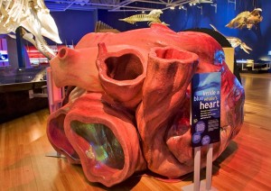
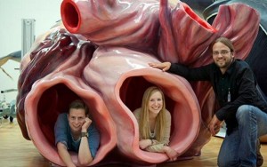
Inside A Blue Whale’s Heart

Many thanks to Bombay Segundo.

Source : Canadian Museum of Nature

Source: @Pickover

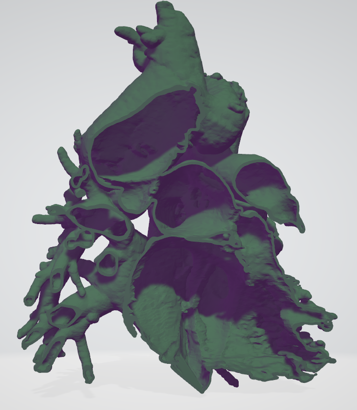
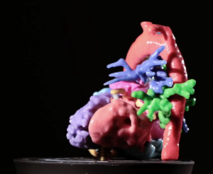
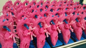
3D Intracardiac Models for Surgical Planning

In multidisciplinary areas, it is very important to be able to meet with the team who can work in harmony.

For his work on 3D intracardiac models on surgical planning,  Thanks to cardiovascular surgeon Dr. Okan Yildiz, we have been informed about and contributed to many pediatric cases and treatments since 2016.

3D Heart_IMAEH

Pulmonary

Link

Anatomical Modeling & 3D Printing Meeting with 4C Medikal

PRINT THYSELF

This sort of procedure is becoming more and more common among doctors and medical researchers. Almost every day, I receive an e-mail from my hospital?s press office describing how yet another colleague is using a 3-D printer to create an intricately realistic surgical model?of a particular patient?s mitral valve, or finger, or optic nerve?to practice on before the actual operation. Surgeons are implanting 3-D-printed stents, prosthetics, and replacement segments of human skull. The exponents of 3-D printing contend that the technology is making manufacturing more democratic; the things we are choosing to print are becoming ever more personal and intimate. This appears to be even more true in medicine: increasingly, what we are printing is ourselves.

Source: Newyorker

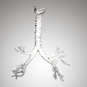
Gigantic Human Organs Made of Glass


Modeling human anatomy. Isn’t it fabulous?

Read more.

Many thanks Sıla Yavuz for this pleasant link.

Trando Med

Trando Med will attend MEDICA 2017 in the Dusseldorf Germany from 13-16 November 2017. The booth is Hall 13 Booth F 9-05

3D Printing for Pediatric Cardiothoracic Surgeons

‘Having worked in product development for the past few years, Dr. Enrique Garcia had seen what 3D printers were capable of and began investigating the possibilities for creating models for pediatric cardiologists to use before an operation. She began by asking surgeons from around the country what they thought of the idea. To say that their response was overwhelmingly positive is an understatement. The value of this idea was immediately apparent.’

‘Pediatric heart surgery is the hardest thing that I can imagine a person doing. A surgeon doesn’t know what he’s going to see until he opens a child?s chest. Every heart is different and every cardiopathy is different,? said Garcia. ?A baby?s heart is the size of a walnut, and surgeons need to go in and move around structures that are as small and thin as a human hair, and they’re doing it with their own two hands. And all of this is occurring against a ticking clock.’

‘Having something in your hands, and being able to turn it any way you want, and to be able to cut and open it up and see the inside; and to be able to physically hold it, to feel it, is something that can?t be replicated on a computer.’

Read More in the source.

Aorta

Instagram

Voksel 3D Surgical Planning with Simpleware -İstanbul

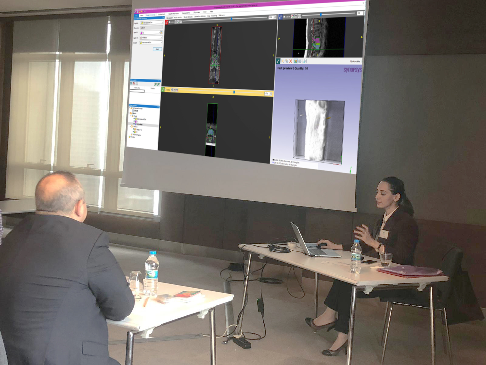
We depicted a live- surgical planning scenario with Prof. Erbil Oğuz and Kerem Girgin in Voksel 3D event. We used Simpleware for image processing, segmentation and designing.

ECVS Conference

It was  sensible showing 3D printed models when i was presenting my study.

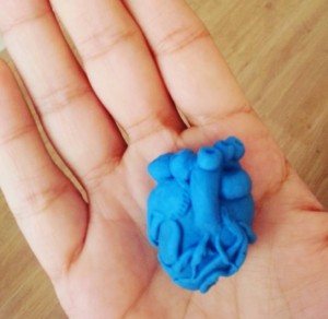
MY TINY DIY HEART

Back to childhood:  I spend my time with play-dough ;)

Link

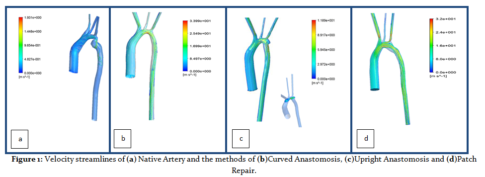
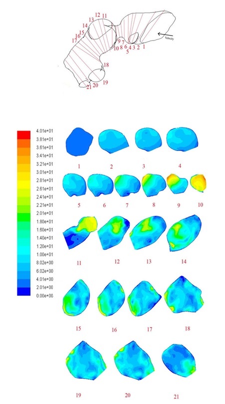
Velocity Contours in Iso-Surfaces for The Re-Stenosed Pulmonary Artery

The entrance lenght parts of the inlet and outlet sections are only used for calculation process.

A 3D Patient-Specific Aorta Model which is Segmented by The Visualisation Toolkit

Wilhelmina Children’s Hospital / Utrecht Sessions – The Netherlands

3 daags kindercardiologie TGA symposium

UMC Utrecht

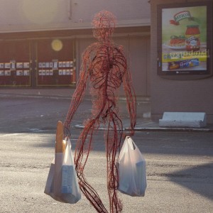
If the human body were deduced to only the circulatory system..

Markos Kay partnered with Jan Kriwol, a Polish photographer who has an interest in optical illusion, in 2012 to begin their project, entitled Human After All. ”The biggest challenge for this project was creating an anatomical character that looked life-like and integrated with the real environment,” Kay tells Creators. “We spent a lot of time experimenting with different postures, and oftentimes we had to exaggerate the posture greatly so that it could translate visually with the deconstructed structure of the circulatory system.”

Source

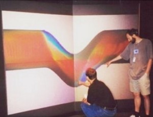
Computational Fluid Dynamics

Link

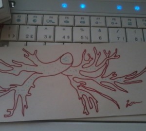
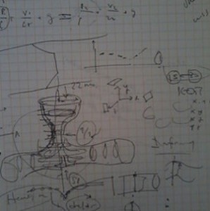
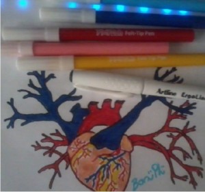
My Lab Drawings – II

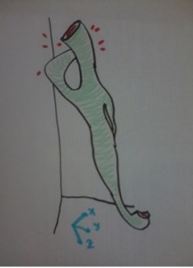
A pulmonary artery. Actually this was the first step of my master thesis.

This will be a collar <3

Link

A Banana with MRI scanning

In the process of learning to create 3D images from MRI and CT datas, bestows you this kind of visual beanos.

Surgical Planning and 3D Printed Hearts

Alistair Phillips, MD, who is the Co-Chair for the American College of Cardiology, Surgeons Section tells about some of the impacts he has personally experienced using 3D printing in surgical settings as his participation in the 3DHEART program:

“The clinical trial is particularly exciting as it targets specific cases in which understanding of the anatomy will greatly enhance the surgical approach. A 3D printed replica of a patient’s heart will be created as part of the inclusion criteria to be in the study.Using 3D printing gave a better understanding of the Hybrid procedure, and allowed us to perform pulmonary valve replacement with a minimally invasive approach avoiding conventional method that required open-heart surgery. After coming to Cedars-Sinai we refined the pre-ventricular approach by utilizing a 3D printed models of patients’ hearts. We were able to simulate the implant into the right ventricular outflow tract.

Every surgeon is different. The education, experience, aptitudes, and attitude we bring to each equally nuanced and varied patient span an almost limitless spectrum and inform how we may utilize 3D printing for the benefit of our patients. The elegance of 3D printing is that it can create the individualized tools spanning this spectrum.

That said, however, what is not negotiable is the veracity of the models that we are receiving. Various materials and their corresponding colouring or rigidity may serve different functions in the hands of different surgeons, but ultimately we must have the utmost confidence in the fidelity of the models we are utilizing for pre-surgical planning. The more realistic the model is both in anatomical and textural preciousness will greatly enhance the application.

In all honesty, I would advise each hospital to start by really understanding the value proposition 3D printing offers across all specialities and, the culture of their institution. The best way to get answers to these very nebulous, complicated, nuanced directives is by retaining an outside vendor to provide as much of the services as possible, from the proverbial soup to nuts.

The excitement around the 3DHEART clinical trial is so great because it is the first organized, large-scale attempt to collect evidence of the efficacies of 3D printing in the practice of medicine and delivery of healthcare, not only in terms of optimized patient outcomes but also with respect to lower costs. If we can get reimbursement for 3D models, it is without a doubt a game-changer in terms of the practice of medicine, and a life-changer for many of our patients.”

Source

ECG

CT scans find clogged arteries in mummies

“We found that heart disease is a serial killer that has been stalking mankind for thousands of years,” says researcher Gregory Thomas. “In the last century, atherosclerotic vascular disease has replaced infectious disease as the leading cause of death across the developed world.”

The researchers performed CT scans of 137 mummies from across four continents and found artery plaque in every single population studied, from preagricultural hunter-gatherers in the Aleutian Islands to the ancient Puebloans of southwestern United States.

Their findings provide an important twist to one?s understanding of atherosclerotic vascular disease, which is the leading cause of death in the developed world: While modern lifestyles can accelerate the development of plaque on arteries, the prevalence of the disease across human history shows it may have a more basic connection to inflammation and aging.

?This is not a disease only of modern circumstance but a basic feature of human aging in all populations,? says Professor Caleb Finch of University of Southern California (USC), a senior author of the study.

?Turns out even a Bronze Age guy from 5,000 years ago had calcified, carotid arteries,? Finch says, referring to Ötzi the Iceman, a natural mummy who lived around 3200 B.C. and was discovered frozen in a glacier in the Italian Alps in 1991.

With Gregory Thomas of Long Beach Memorial, Finch was part of a team that previously showed Egyptian mummies had calcified patches on their arteries indicative of advanced atherosclerosis (from the Greek ?athero,? meaning ?gruel,? and ?scler,? meaning ?hard?).

But ancient Egyptians tended to mummify only royalty or those who had privileged lives. The new study led by Thomas and Randall Thompson of Saint Luke?s Mid America Heart Institute examined mummies from four drastically different climates and diets?and from cultures that mummified regular people, including ancient Peruvians, Ancestral Puebloans, the Unangans of the Aleutian Islands, and ancient Egyptians.

?Our research shows that we are all at risk for atherosclerosis, the disease that causes heart attacks and strokes?all races, diets, and lifestyles,? says Thomas.

?Because of this we all need to be cautious of our diet, weight, and exercise to minimize its impact. The data gathered about individuals from the prehistoric cultures of ancient Peru and the Native Americans living along the Colorado River and the Unangan of the Aleutian Islands is forcing us to think outside the box and look for other factors that may cause heart disease.?

Overall, the researchers found probable or definite atherosclerosis in 34 percent of the mummies studied, with calcification of arteries more pronounced in the mummies that were older at the time of death. Atherosclerosis was equally common in mummies identified as male or female.

?We found that heart disease is a serial killer that has been stalking mankind for thousands of years,? Thompson says. ?In the last century, atherosclerotic vascular disease has replaced infectious disease as the leading cause of death across the developed world.

?A common assumption is that the rise in levels of atherosclerosis is predominantly lifestyle-related, and that if modern humans could emulate preindustrial or even preagricultural lifestyles, that atherosclerosis, or at least its clinical manifestations, would be avoided.

?Our findings seem to cast doubt on that assumption, and at the very least, we think they suggest that our understanding of the causes of atherosclerosis is incomplete and that it might be somehow inherent to the process of human aging,? he adds.

The researchers will next seek to biopsy ancient mummies to get a better understanding of the role chronic infection, inflammation, and genetics play in promoting the prevalence of atherosclerosis.

?Atherosclerosis starts very early in life. In the United States, most kids have little bumps on their arteries. Even stillbirths have little tiny nests of inflammatory cells. But environmental factors can accelerate this process,? Finch says, pointing to studies that show larger plaque buildup in children exposed to household tobacco smoking or who are obese.

Source: University of Southern California

My Lab Drawings – I

Beliz and me in the lab.

His name is tramp carotid :)

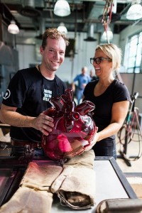
Handmade Glass Anatomical Models by Farlow’s Scientific Glassblowing

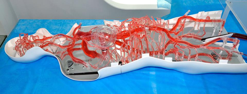
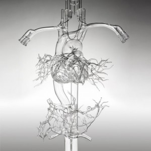
Gary Farlow can make art out of arteries. He and his team of 10 at Farlow?s Scientific Glassblowing are able to transform the body?s vasculature?and nearly all of its other parts?into an ornate borosilicate glass sculpture, from the heart?s ventricles to the brain?s circle of Willis. ?We do almost every part of the body,? Farlow says. ?It can take a pretty artistic mind to make some of these things.? With the help of cardiologists, the team creates custom see-through systems for science and medical training.

Their anatomically correct models can be designed to simulate blood flow, teach placement of catheters and angioplasty devices, or simply test or demo new surgical gizmos. Individual arteries, veins, and capillaries are shaped and fused together, one at a time. Ground-glass joints are added at the exposed ends so a head, say, can be connected to the carotid arteries should customers want to expand their model. A full-body setup could cost $25,000, so don?t get any bright ideas about using one as a brandy decanter.

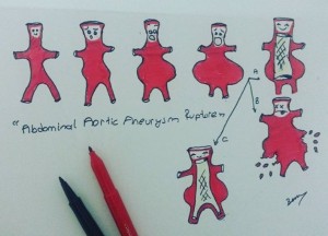
Aorta Aneurysm

Knee Anatomy Segmentation

I tried to show the knee anatomy with the MRI dataset of 3D Slicer (Harvard Medical School /Brigham and Women’s Hospital / Surgical Planning Laboratory).

Video: Knee Anatomy